Endosseous Implant Failure Influenced by Crown Cementation: A Clinical Case Report
Ricardo Gapski, DDS, BDS, MS1/Neil Neugeboren, DDS1/Alan Z. Pomeranz, DMD, MMSc1/Marc W. Reissner, DDS1
Implant dentistry has developed predictable treatment outcomes. Nevertheless, there are multiple rea-sons for implant failure. This case report documents a previously unreported type of implant failurethat occurred 1 month after crown cementation. The implant failure is believed to be associated withretained excess subgingival cement. INT J ORAL MAXILLOFAC IMPLANTS 2008;23:943–946
Key words: cement, complication, dental implant, implant loss
Titanium endosseous dental implants have been to prosthetic reasons is scarce. This case report
increasingly utilized over the past few decades.1
relates to a prosthetic-related implant complication
Successful outcomes can be expec ted when
that resulted in early implant failure.
implants are placed in bone of good quality andquantity and when proper surgical protocol is fol-lowed.2 Although dental implants are considered a
very successful mode of therapy, many factors havebeen associated with the failure of dental implants.3
A 31-year-old Hispanic woman presented to the
Complicating factors can be divided into the follow-
authors’ periodontal office reporting mobility of the
ing categories: surgery-related implant loss; bone
maxillary right lateral incisor. The medical history of
loss; peri-implant soft tissue disease; mechanical
the patient was noncontributor y. The patient
problems; and esthetic/phonetic results.4
reported previous orthodontic therapy for 3 years. A
In terms of biological implant failure, contributing
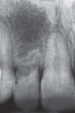
periapical radiograph revealed severe root resorp-
factors reported in the literature include implant
tion (Fig 1). The treatment plan was to extract the
length and diameter,5 body design,6 smoking,1,5
maxillary right lateral incisor with immediate place-
implant location,7 bone quality,8 peri-implantitis,9
ment of an endosseous dental implant. After local
and others. In terms of mechanical implant failure,
anesthesia was obtained, the maxillary right lateral
several investigations have evaluated the most com-
incisor was atraumatically extracted. The surgical site
mon prosthetic complications associated with dental
revealed an adequate amount of alveolar bone for
implants. Overall, the majority of these studies focus
immediate implant placement. The buccal alveolar
on problems associated with the suprastructural
bone was intact, and no signs of pathology or bone
components and the function/esthetics of the pros-
resorption beyond the socket of the remaining tooth
thesis. Examples of such complications are abutment
fractures and loosening,10 prosthesis fracture,11,12
Subsequently, a narrow, internal platform, parallel-
prosthesis retention and comfort,13 and patient satis-
walled endosseous implant was inserted (3.25 ϫ 11.5
faction.13 The literature on early implant failure due
mm; Biomet 3i, Palm Beach Gardens, FL) using a surgicaltemplate (Fig 2). At the same visit, the healing abut-ment was inserted (3.4 ϫ 4 mm) and a provisionalremovable partial denture was delivered. Postoperativemedication included amoxicillin 500 mg every 8 hours
for 10 days, chlorhexidine 0.12% every 12 hours for 7days, and an acetaminophen/hydrocodone-based anal-
Correspondence to: Dr Ricardo Gapski, 10200 East Girard
gesic as needed for pain. The implant was allowed to
Avenue, Building A, Suite 209, Denver, CO 80231. Fax: +303 6956915. E-mail: [email protected]
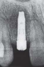
heal for 4 months (Fig 3). Then, a reverse torque of 20
The International Journal of Oral & Maxillofacial Implants
COPYRIGHT 2008 BY QUINTESSENCE PUBLISHING CO, INC. PRINTING OF THIS DOCUMENT IS RESTRICTED TO PERSONAL USE ONLY. NO
PART OF THIS ARTICLE MAY BE REPRODUCED OR TRANSMITTED IN ANY FORM WITHOUT WRITTEN PERMISSION FROM THE PUBLISHER
graph. Note severe resorption of tooth inthe maxillary right lateral incisor.
strating uneventful healing 4 months afterimplant placement.
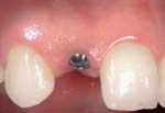
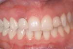
imately 1 month after crown cementationwas performed. Note the erythematous andcyanotic tissues around the implant. A 9-mm peri-implant pocket with suppurationwas detected on the distal aspect of theimplant.
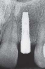
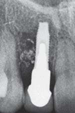
mately 1 month after crown cementation. Note the radiopaque material at the distalaspect of the implant in combination withextensive bone loss.
Ncm was utilized to ensure the implant was osseointe-
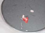
extensive bone loss (Fig 6). It was decided to remove
grated and a follow-up radiograph was obtained (Fig 4).
the crown and prosthetic abutment and re-insert the
The peri-implant sulcus was within normal range.
healing abutment and provisional partial denture
The patient returned to the periodontal office 1
prior to exploration of the site. After local anesthesia
month after final cementation of the implant pros-
was obtained, the area was surgically explored,
thesis reporting soreness and swelling in the area
revealing extensive bone loss distal and buccal to
(Fig 5). Clinically, a 9-mm pocket with suppuration
the implant (Figs 7a and 7b). There was a mixture
was present on the distal aspect of the implant, while
resembling granulation material and temporary
a shallow sulcus was present on the mesial aspect of
cement involving up to 70% of the implant length.
the maxillary right canine. A radiograph at the same
The implant was surgically removed, and guided
appointment revealed radiopaque material at the
bone regeneration was performed at the site for a
distal aspect of the implant in combination with
COPYRIGHT 2008 BY QUINTESSENCE PUBLISHING CO, INC. PRINTING OF THIS DOCUMENT IS RESTRICTED TO PERSONAL USE ONLY. NO
PART OF THIS ARTICLE MAY BE REPRODUCED OR TRANSMITTED IN ANY FORM WITHOUT WRITTEN PERMISSION FROM THE PUBLISHER
defect revealed extensive bone loss at thedistal and buccal aspects of the implantand material resembling temporary cementat the thread of the implant. (b) A mixture ofgranulation tissue with large amount ofmaterial resembling temporary cement wasremoved from the defect.
retrieval and excess cement removal can be experi-enced with cemented restorations.17 When properly
Numerous studies show that abutment loosening
restored, the intracrevicular position of the restora-
constitutes one of the known implant postsurgery
tion margin does not appear to adversely affect peri-
complications requiring clinical intervention.4 A
implant health and stability.18 However, it can be
review of the literature demonstrated that abutment
speculated that excess cement is more difficult to
loosening is the most common prosthetic complica-
remove or identify when implants are restored with
tion in implant dentistry (2% to 45% depending on
deep subgingival margins. These situations are more
the study and type of prosthesis).4 In a prospective
commonly seen in anterior restorations, where
preclinical study, 27% of loosened screws were pre-
esthetic demands are higher. In such cases, the mar-
sent with use of screwed abutments, in comparison
gins are usually placed further subgingivally, leading
to no abutment loosening with cemented restora-
to an increased risk of leaving additional cement in
tions.14 The authors speculated that screwed abut-
the peri-implant tissues. In a recent 8-year private
ments are often submitted to nonaxial loads that
practice study, the authors did not notice different
determine screw and abutment loosening.14 Screw
complication rates for cemented and screw-retained
loosening not only becomes an inconvenience to
prostheses.10 However, the authors recommended
clinicians and patients due to the increase in mainte-
screw-retained prostheses in the esthetic zone to
nance, but also there are biological detrimental
avoid problems associated with excess cement irri-
effects in the surrounding tissues when this condi-
tion occurs. An in vivo study has demonstrated an
One of the reasons for such a complication possi-
increase in expression of vascular endothelial growth
bly relates to the supracrestal soft tissue micro-
factor and microvessel density (markers of inflamma-
anatomy around dental implants. In contrast to nat-
tion) in loosely screwed abutments compared to
ural teeth, implants do not develop perpendicular
screw-tight and cement-retained restorations.15 In
fiber attachment.19,20 Instead, the gingival connec-
addition, microbial leak age through the gap
tive tissue fibers are closely adapted to the titanium
between the suprastructure and the abutment plays
layer but in an orientation approximately parallel to
an important role in the bacterial colonization of the
the implant surface.20 This anatomic condition may
internal part of screw-retained crowns and partial
not provide enough protection if excess cement is
pushed into the peri-implant sulcus. In fact, probing
Despite all the advantages related to cement-
measurements around healthy osseointegrated oral
retained implant restorations, some disadvantages
implants and teeth differ.21 Histologic studies have
can be clearly seen. For instance, difficult prosthesis
demonstrated that a probe has a tendency to pene-
The International Journal of Oral & Maxillofacial Implants
COPYRIGHT 2008 BY QUINTESSENCE PUBLISHING CO, INC. PRINTING OF THIS DOCUMENT IS RESTRICTED TO PERSONAL USE ONLY. NO
PART OF THIS ARTICLE MAY BE REPRODUCED OR TRANSMITTED IN ANY FORM WITHOUT WRITTEN PERMISSION FROM THE PUBLISHER
trate deeper into the peri-implant tissues compared
7. Hutton JE, Heath MR, Chai JY, et al. Factors related to success
to the counterpart teeth.21–23 In addition, it has been
and failure rates at 3-year follow-up in a multicenter study of
demonstrated that peri-implant probing depth mea-
overdentures supported by Brånemark implants. Int J OralMaxillofac Implants 1995;10:33–42.
surements are more sensitive to force variation than
8. Khang W, Feldman S, Hawley CE, et al. A multi-center study
periodontal pocket probing.22 Hence, it could be fur-
comparing dual acid-etched and machined-surfaced implants
ther speculated that implants may be more sensitive
in various bone qualities. J Periodontol 2001;72:1384–1390.
to excess cement pressed into the peri-implant tis-
9. Esposito M, Hirsch JM, Lekholm U, et al. Biological factors con-
sue than tissue around natural teeth.
tributing to failures of osseointegrated oral implants. (II). Etiopathogenesis. Eur J Oral Sci 1998;106:721–764.
This is the first case report demonstrating that
10. Nedir R, Bischof M, Szmukler-Moncler S, et al. Prosthetic com-
excess cement can lead to severe clinical conse-
plications with dental implants: From an up-to-8-year experi-
quences, including implant failure. However, it is diffi-
ence in private practice. Int J Oral Maxillofac Implants 2006;
cult to be certain of the cause-effect cited in this
report; other factors should be considered. First,
11. Krennmair G, Piehslinger E, Wagner H. Status of teeth adjacent
to single-tooth implants. Int J Prosthodont 2003;16:524–548.
there were no apparent signs of peri-implant pathol-
12. Gothberg C, Bergendal T, Magnusson T. Complications after
ogy prior to the insertion of the crown. Second, the
treatment with implant-supported fixed prostheses: A retro-
crown was not in occlusal trauma, which could justify
spective study. Int J Prosthodont 2003;16:201–207.
the severe bone loss. Finally, there was a large
13. Naert I, Alsaadi G, Quirynen M. Prosthetic aspects and patient
amount of temporary cement in the vertical defect
satisfaction with two-implant-retained mandibular overden-tures: A 10-year randomized clinical study. Int J Prosthodont
around the implant. It is imperative to note that it is
difficult to speculate whether local factors such as an
14. Assenza B, Scarano A, Leghissa G, et al. Screw- vs cement-
undermining bone fenestration influenced the sever-
implant-retained restorations: An experimental study in the
ity of the infection. Further controlled studies are
Beagle. Part 1. Screw and abutment loosening. J Oral Implan-
necessary to unravel all possible variables associated
15. Assenza B, Artese L, Scarano A, et al. Screw vs cement-implant-
with excess cement and implant failure.
retained restorations: An experimental study in the beagle. Part 2. Immunohistochemical evaluation of the peri-implanttissues. J Oral Implantol 2006;32:1–7.
16. Keller W, Bragger U, Mombelli A. Peri-implant microflora of
implants with cemented and screw retained suprastructures. Clin Oral Implants Res 1998;9:209–217.
Cementation of implant-supported crowns is com-
17. Rajan M, Gunaseelan R. Fabrication of a cement- and screw-
mon in the esthetic zone. It is recommended that the
retained implant prosthesis. J Prosthet Dent 2004;92:578–580.
restorative dentist be especially vigilant to remove
18. Giannopoulou C, Bernard JP, Buser D, et al. Effect of intracrevic-
excess subgingival cement following cementation of
ular restoration margins on peri-implant health: Clinical, bio-
the crowns to avoid the potential for implant failure.
chemical, and microbiologic findings around estheticimplants up to 9 years. Int J Oral Maxillofac Implants 2003;
The biologic process by which the cement is found
within the intraosseous defect requires further study.
19. Cochran DL, Hermann JS, Schenk RK, et al. Biologic width
around titanium implants. A histometric analysis of theimplanto-gingival junction around unloaded and loaded non-
submerged implants in the canine mandible. J Periodontol1997;68:186–198.
20. Listgarten MA, Buser D, Steinemann SG, et al. Light and trans-
1. Schwartz-Arad D, Samet N, Mamlider A. Smoking and compli-
mission electron microscopy of the intact interfaces between
cations of endosseous dental implants. J Periodontol 2002;73:
non-submerged titanium-coated epoxy resin implants and
bone or gingiva. J Dent Res 1992;71:364–371.
2. Brånemark P-I, Hansson BO, Adell R, et al. Osseointegrated
21. Schou S, Holmstrup P, Stoltze K, et al. Probing around implants
implants in the treatment of the edentulous jaw. Experience
and teeth with healthy or inflamed peri-implant mucosa/gin-
from a 10-year period. Scand J Plast Reconstr Surg 1977;
giva. A histologic comparison in cynomolgus monkeys
(Macaca fascicularis). Clin Oral Implants Res 2002;13:113–126.
3. Tolstunov L. Dental implant success-failure analysis: A concept
22. Mombelli A, Muhle T, Bragger U, et al. Comparison of peri-
of implant vulnerability. Implant Dent 2006;15:341–346.
odontal and peri-implant probing by depth-force pattern
4. Goodacre CJ, Bernal G, Rungcharassaeng K, et al. Clinical com-
analysis. Clin Oral Implants Res 1997;8:448–454.
plications with implants and implant prostheses. J Prosthet
23. Ericsson I, Lindhe J. Probing depth at implants and teeth. An
experimental study in the dog. J Clin Periodontol 1993;
5. Bain CA, Moy PK. The association between the failure of dental
implants and cigarette smoking. Int J Oral Maxillofac Implants1993;8:609–615.
6. Karoussis IK, Bragger U, Salvi GE, et al. Effect of implant design
on survival and success rates of titanium oral implants: A 10-year prospective cohort study of the ITI Dental Implant Sys-tem. Clin Oral Implants Res 2004;15:8–17.
COPYRIGHT 2008 BY QUINTESSENCE PUBLISHING CO, INC. PRINTING OF THIS DOCUMENT IS RESTRICTED TO PERSONAL USE ONLY. NO
PART OF THIS ARTICLE MAY BE REPRODUCED OR TRANSMITTED IN ANY FORM WITHOUT WRITTEN PERMISSION FROM THE PUBLISHER
A PUBLICATION OF THE VIRGINIA STATE BEEKEEPERS’ ASSOCIATION New Foulbrood Treatment Fall Meeting Highlights FDA approves Tylan Soluble® for honey bees Notes from VSBA meeting in Weyers Cave American beekeepers will soon have a new antibiotic with About 125 beekeepers from Virginia and neighboring states which to protect their colonies from American foulbrood attended t
Sustained improvement in a patient with young onset Parkinson’sdisease after the arrival of a pet dogReceived: 14 January 2010 / Revised: 11 February 2010 / Accepted: 17 February 2010 / Published online: 16 March 2010Ó Springer-Verlag 2010III: 8/108). However, despite domperidone, she experi-enced continuous nausea, anorexia and fatigue, whichMuch research has been carried out into pharmacol
 graph. Note severe resorption of tooth inthe maxillary right lateral incisor.
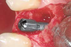
 defect revealed extensive bone loss at thedistal and buccal aspects of the implantand material resembling temporary cementat the thread of the implant. (b) A mixture ofgranulation tissue with large amount ofmaterial resembling temporary cement wasremoved from the defect.
retrieval and excess cement removal can be experi-enced with cemented restorations.17 When properly
Numerous studies show that abutment loosening
restored, the intracrevicular position of the restora-
constitutes one of the known implant postsurgery
tion margin does not appear to adversely affect peri-
complications requiring clinical intervention.4 A
implant health and stability.18 However, it can be
review of the literature demonstrated that abutment
speculated that excess cement is more difficult to
loosening is the most common prosthetic complica-
remove or identify when implants are restored with
tion in implant dentistry (2% to 45% depending on
deep subgingival margins. These situations are more
the study and type of prosthesis).4 In a prospective
commonly seen in anterior restorations, where
preclinical study, 27% of loosened screws were pre-
esthetic demands are higher. In such cases, the mar-
sent with use of screwed abutments, in comparison
gins are usually placed further subgingivally, leading
to no abutment loosening with cemented restora-
to an increased risk of leaving additional cement in
tions.14 The authors speculated that screwed abut-
the peri-implant tissues. In a recent 8-year private
ments are often submitted to nonaxial loads that
practice study, the authors did not notice different
determine screw and abutment loosening.14 Screw
complication rates for cemented and screw-retained
loosening not only becomes an inconvenience to
prostheses.10 However, the authors recommended
clinicians and patients due to the increase in mainte-
screw-retained prostheses in the esthetic zone to
nance, but also there are biological detrimental
avoid problems associated with excess cement irri-
effects in the surrounding tissues when this condi-
tion occurs. An in vivo study has demonstrated an
One of the reasons for such a complication possi-
increase in expression of vascular endothelial growth
bly relates to the supracrestal soft tissue micro-
factor and microvessel density (markers of inflamma-
anatomy around dental implants. In contrast to nat-
tion) in loosely screwed abutments compared to
ural teeth, implants do not develop perpendicular
screw-tight and cement-retained restorations.15 In
fiber attachment.19,20 Instead, the gingival connec-
addition, microbial leak age through the gap
tive tissue fibers are closely adapted to the titanium
between the suprastructure and the abutment plays
layer but in an orientation approximately parallel to
an important role in the bacterial colonization of the
the implant surface.20 This anatomic condition may
internal part of screw-retained crowns and partial
not provide enough protection if excess cement is
pushed into the peri-implant sulcus. In fact, probing
Despite all the advantages related to cement-
measurements around healthy osseointegrated oral
retained implant restorations, some disadvantages
implants and teeth differ.21 Histologic studies have
can be clearly seen. For instance, difficult prosthesis
demonstrated that a probe has a tendency to pene-
The International Journal of Oral & Maxillofacial Implants
COPYRIGHT 2008 BY QUINTESSENCE PUBLISHING CO, INC. PRINTING OF THIS DOCUMENT IS RESTRICTED TO PERSONAL USE ONLY. NO
PART OF THIS ARTICLE MAY BE REPRODUCED OR TRANSMITTED IN ANY FORM WITHOUT WRITTEN PERMISSION FROM THE PUBLISHER
trate deeper into the peri-implant tissues compared
7. Hutton JE, Heath MR, Chai JY, et al. Factors related to success
to the counterpart teeth.21–23 In addition, it has been
and failure rates at 3-year follow-up in a multicenter study of
demonstrated that peri-implant probing depth mea-
overdentures supported by Brånemark implants. Int J OralMaxillofac Implants 1995;10:33–42.
defect revealed extensive bone loss at thedistal and buccal aspects of the implantand material resembling temporary cementat the thread of the implant. (b) A mixture ofgranulation tissue with large amount ofmaterial resembling temporary cement wasremoved from the defect.
retrieval and excess cement removal can be experi-enced with cemented restorations.17 When properly
Numerous studies show that abutment loosening
restored, the intracrevicular position of the restora-
constitutes one of the known implant postsurgery
tion margin does not appear to adversely affect peri-
complications requiring clinical intervention.4 A
implant health and stability.18 However, it can be
review of the literature demonstrated that abutment
speculated that excess cement is more difficult to
loosening is the most common prosthetic complica-
remove or identify when implants are restored with
tion in implant dentistry (2% to 45% depending on
deep subgingival margins. These situations are more
the study and type of prosthesis).4 In a prospective
commonly seen in anterior restorations, where
preclinical study, 27% of loosened screws were pre-
esthetic demands are higher. In such cases, the mar-
sent with use of screwed abutments, in comparison
gins are usually placed further subgingivally, leading
to no abutment loosening with cemented restora-
to an increased risk of leaving additional cement in
tions.14 The authors speculated that screwed abut-
the peri-implant tissues. In a recent 8-year private
ments are often submitted to nonaxial loads that
practice study, the authors did not notice different
determine screw and abutment loosening.14 Screw
complication rates for cemented and screw-retained
loosening not only becomes an inconvenience to
prostheses.10 However, the authors recommended
clinicians and patients due to the increase in mainte-
screw-retained prostheses in the esthetic zone to
nance, but also there are biological detrimental
avoid problems associated with excess cement irri-
effects in the surrounding tissues when this condi-
tion occurs. An in vivo study has demonstrated an
One of the reasons for such a complication possi-
increase in expression of vascular endothelial growth
bly relates to the supracrestal soft tissue micro-
factor and microvessel density (markers of inflamma-
anatomy around dental implants. In contrast to nat-
tion) in loosely screwed abutments compared to
ural teeth, implants do not develop perpendicular
screw-tight and cement-retained restorations.15 In
fiber attachment.19,20 Instead, the gingival connec-
addition, microbial leak age through the gap
tive tissue fibers are closely adapted to the titanium
between the suprastructure and the abutment plays
layer but in an orientation approximately parallel to
an important role in the bacterial colonization of the
the implant surface.20 This anatomic condition may
internal part of screw-retained crowns and partial
not provide enough protection if excess cement is
pushed into the peri-implant sulcus. In fact, probing
Despite all the advantages related to cement-
measurements around healthy osseointegrated oral
retained implant restorations, some disadvantages
implants and teeth differ.21 Histologic studies have
can be clearly seen. For instance, difficult prosthesis
demonstrated that a probe has a tendency to pene-
The International Journal of Oral & Maxillofacial Implants
COPYRIGHT 2008 BY QUINTESSENCE PUBLISHING CO, INC. PRINTING OF THIS DOCUMENT IS RESTRICTED TO PERSONAL USE ONLY. NO
PART OF THIS ARTICLE MAY BE REPRODUCED OR TRANSMITTED IN ANY FORM WITHOUT WRITTEN PERMISSION FROM THE PUBLISHER
trate deeper into the peri-implant tissues compared
7. Hutton JE, Heath MR, Chai JY, et al. Factors related to success
to the counterpart teeth.21–23 In addition, it has been
and failure rates at 3-year follow-up in a multicenter study of
demonstrated that peri-implant probing depth mea-
overdentures supported by Brånemark implants. Int J OralMaxillofac Implants 1995;10:33–42.