Doi:10.1016/j.jocn.2004.03.01

Journal of Clinical Neuroscience (2005) 12(3), 221–2300967-5868/$ - see front matter ª 2004 Elsevier Ltd. All rights reserved.
doi:10.1016/j.jocn.2004.03.011 Imaginem oblivionis: the prospects of neuroimaging for earlydetection of Alzheimer’s diseaseq,qq Victor L. Villemagne1,2,3 MD, C.C. Rowe1,4 MD FRACP, S. Macfarlane2,3 FRANZCP, K.E. Novakovic1,3 BSC,C.L. Masters2,3 MD FRCPA 1Department of Nuclear Medicine, Centre for PET, Austin Hospital, Melbourne, Vic., Australia, 2Mental Health Research Institute, Parkville, Vic., Australia,3Department of Pathology, University of Melbourne, Vic., Australia, 4Department of Medicine, University of Melbourne, Vic., Australia Summary Alzheimer’s disease (AD) is a progressive neurodegenerative disorder characterised by the gradual onset of dementia. Thepathological hallmarks of the disease are Ab amyloid plaques, neurofibrillary tangles, and reactive gliosis.
Current diagnosis of AD is made by clinical, neuropsychologic, and neuroimaging assessments. Routine structural neuroimaging evaluation is based on non-specific features such as atrophy, a late feature in the progression of the disease, hence the crucial importance of developingnew approaches for early and specific recognition at the prodromal stages of AD.
Functional neuroimaging techniques such as positron emission tomography (PET) and single photon emission computed tomography (SPECT) could prove to be valuable in the differential diagnosis of AD, as well as in assessing prognosis. With the advent of new therapeuticstrategies aimed at reducing the Ab amyloid burden in the brain, there is increasing interest in the development of PET and SPECT radioligandsthat will permit the assessment of Ab amyloid burden in vivo. From this, the prospect of specific preclinical diagnosis arises, possibly inconjuction with other related Ab biomarkers in plasma and CSF.
ª 2004 Elsevier Ltd. All rights reserved.
Keywords: functional neuroimaging, Alzheimer’s disease, Ab amyloid, PET, SPECT criteria followed in most research studies are those proposed bythe National Institute of Neurological and Communicative Alzheimer’s disease (AD) is a progressive neurodegenerative Disorders and Stroke-Alzheimer’s Disease and Related Disorders disorder characterised by the gradual onset of dementia,1 leading invariably to death, usually between 7 and 10 years after diag- nosis. Age is the major risk factor. AD is the leading cause of A variable period of prodromal decline in cognition of up to dementia in the elderly and affects about 5% of individuals at age five years usually precedes the formal diagnosis of AD. This 65, with its prevalence increasing exponentially with each suc- stage, known as Minimal Cognitive Impairment (MCI), is char- cessive decade, with 10–15% and 20–30% of 75- and 85-year olds acterized by a relatively isolated impairment in long term memory and may also be accompanied by impairments of working mem-ory (Table 2). These deficits presumably relate to damage to the medial temporal lobe and/or specific prefrontal–temporal lobe circuits. About 40–60% of carefully characterized subjects with The clinical diagnosis of AD is currently based on progressive MCI will subsequently progress to meet criteria for AD over a 3– memory impairment and decline in at least one other cognitive domain, and by excluding other diseases such as frontotemporal dementia, dementia with Lewy-bodies (DLB), stroke, brain tu-mour, normal pressure hydrocephalus or depression, that might While mutations of the PS-1, PS-2,11 and amyloid precursor protein (APP) genes – resulting in increased production and ele- The clinical diagnostic accuracy for AD depends on the stage vated plasma levels of amyloid-b protein – on chromosomes 1, 14, of disease and can exceed 90% in academic settings in mid or late and 21, respectively, have been associated with the rare form of stages.4 Diagnostic criteria for AD have been proposed within familial AD, the only consistent marker for the late-onset non- both the DSM5 and ICD classification systems.6 However, the familial form of dementia is the apolipoprotein e allele on chro-mosome 19.12 This refers specifically to the e4 allele, though it is absent in approximately 30–40% of patients with AD and present Supported in part by grants from the National Health and Medical in about 30% of healthy subjects.13 On the other hand, the e3 Research Council of Australia, Prana Biotechnology and Schering AG.
qq allele is believed to represent no increased or decreased risk, while Quo pacto dicam imaginem oblivionis teneri memoria mea, non ipsam the e2 allele may confer some protection.14 oblivionem, cum eam memini? (How can I assert that the image of oblivionis retained by my memory, and not oblivion itself, when I remember it?) St Augustine. Confessiones. Liber X, Caput 16.
By means of clinical, laboratory, and imaging evidence only a provisional diagnosis of either possible or probable AD can bemade in living subjects. In the absence of biologic markers, direct Correspondence to: Dr. Victor L. Villemagne MD, Department of Nuclear pathologic examination of brain tissue derived from either biopsy Medicine, Centre for PET, Austin Health, 145 Studley Road, Heidelberg, Vic.
3084, Australia. Tel.: +61-3-9496-3321; Fax: +61-3-9458-5023; or autopsy remains the only definitive method for establishing a NINCDS-ARDA criteria defining probable AD are most easily identified in the hippocampus. NFTs are notspecific to AD and are found in a variety of other neurodegen-  Dementia established by clinical examination and documented by the erative conditions such as Down's syndrome, subacute sclerosing mini-mental test (MMSE), Blessed Dementia scale, or similar examination, panencephalitis, Hallervorden-Spatz disease, Parkinson dementia and confirmed by neuropsychological tests; complex, and dementia pugilistica.15;20 A number of in vitro and in  Deficits in two or more areas of cognition; Progressive worsening of memory and other cognitive functions; vivo studies have shown Ab protein to be directly toxic to neu- rons, leading to the aggregation and secondary phosphorylation of  Onset between ages of 40 and 90, most often after age 65;  Absence of other systemic disorders or brain diseases that in and of themselves could account for the progressive deficits in memory andcognition.
Amyloid plaques are extracellular aggregations of amyloid proteinof about 50–100 lm in diameter intimately surrounded by dys- trophic axons and dendrites, reactive astrocytes, and activatedmicroglia. Though mainly located in the amygdala and hippo-  Memory complaint (subjective memory impairment) campus, they are present throughout the cortex.19 The primary  Normal activities of daily living (ADL) component of the plaque is the amyloid b protein (Ab), a 40–43 amino acid peptide (4 kDa) product of the proteolytic cleavage of APP by b- or c-secretases.22;23 The peptide is referred to as “b”amyloid due to its secondary structure of b-pleated sheets. Ab isnot only found within senile plaques, but is also present around The typical macroscopic picture is gross cortical atrophy.
cortical arterioles as a congophilic angiopathy.
Microscopically, there is widespread cellular degeneration and Although it is as yet unproven whether the increased produc- neuronal loss that affects primarily the outer three layers of the tion, precipitation, and progressive deposition of Ab is causative cerebral cortex, initially affecting more the temporal and frontal in terms of the pathogenesis of AD,24 or merely an epiphenome- cortical regions subserving cognition than the parietal and non of the disease process, the Ab theory of AD is the dominant occipital cortices. These changes are accompanied by reactive etiologic paradigm at this time. Genetic mutations within the APP gliosis and by the presence of the pathological hallmarks of the gene cause rare cases of early-onset familial AD, and other disease, intracellular neurofibrillary tangles (NFTs) and extracel- causative mutations within genes associated with the c secretase complex (presenilin 1, 2) are the most convincing evidence thatAb production is the causative factor at the centre of AD patho-genesis. While the exact mechanism by which Ab might produce cell death is controversial, it is believed that a toxic oxidative Neurofibrillary tangles are intraneuronal bundles of paired helical interaction between Ab and various metal species leads to lipid filaments. The main structural component of NFTs is a normal damaging and/or oxidative response with free radical production constituent of cellular microtubules but present in AD in an leading to progressive disruption of neuronal function and ulti- abnormally phosphorylated form, known as Tau protein.18;19 They Neurofibrillary tanglesand neuropil threads Schematical representation of the cascade of events leading to Ab deposition and oxidative injury.
Journal of Clinical Neuroscience (2005) 12(3), 221–230 ª 2004 Elsevier Ltd. All rights reserved.
Structural neuroimaging techniques, such as computed tomogra- phy (CT) and magnetic resonance imaging (MRI), are routinely used in the clinical evaluation of AD patients.
Widespread cortical atrophy with a thinning of medial tem- poral lobe structures are the most consistent structural neuroim-aging findings associated with AD,26 though not pathognomonicof the disease because there is overlap with “normal” aging.
tissue of AD patients can be obtained non-invasively through CT is mainly used to exclude other treatable causes of proton MRS.47 Some MRS studies have shown regional decreases dementia.27 Studies reporting AD-related changes found that in NAA in patients with AD in temporal and parietal cortices,48–51 whilst all dementia groups tended to display greater medial tem- while also demonstrating a positive correlation between the de- poral lobe (MTL) atrophy on CT compared to the depressed pa- gree of NAA reductions and disease severity by neuropathologic tient group, MTL atrophy was unable to discriminate between criteria.52;53 MRS has also been applied to monitor response to therapeutic interventions in AD.54;55 Patients with AD showed MRI has been used to examine atrophy of the entorhinal, significantly higher mean diffusibility in hippocampus, cingulate, perirhinal, and temporal cortices in patients with early AD,29 temporal, and parietal white matter than a control group using where the severity of volume loss was correlated with disease diffusion weighted MR imaging,56 while diffusion tensor MR severity. These regions were also found to be reduced in both MCI imaging has shown diffuse reduction in white matter integrity in and AD compared to controls in AD.30;31 Both conditions were also significantly associated with cortical grey matter loss and Both PET and SPECT are molecular imaging techniques that ventricular enlargement. Other studies32 suggested that measure- use radiolabelled tracers to evaluate biological processes in ments of hippocampal changes were more practical and useful.
vivo43;58;59 (Table 3). These techniques already play a role in the Hippocampal and parahippocampal volume loss was signifi- differential diagnosis of AD from other dementing conditions such cantly greater in patients with AD than in patients with DLB33 as vascular dementia, frontotemporal dementia, DLB, and consistent with the relative preservation of memory that is seen in PET is a sensitive molecular imaging technique that allows in Volumetric changes on MRI are entirely consistent with the vivo quantification in absolute values of the concentration of a patterns of neuropathological progression in AD, with damage to radiotracer, where either the radiotracer bears the same bio- the medial temporal lobe and association cortex accounting for the chemical structure or is an analog, or is a substrate of the chemical classical patterns of cognitive impairment that are commonly process being evaluated, allowing the in vivo assessment of the observed in AD.34;35 Substantial neuronal loss occurred by the molecular process at their sites of action.58 With a theoretical time atrophy is detectable by MRI.36;37 Furthermore, the absence spatial resolution of 3–4 mm, which in practice translates to 5–6 of cortical atrophy or medial temporal lobe changes is not suffi- mm, and an exquisite sensitivity (detecting concentrations in the picomolar range), the technique may permit detection of disease The fact that structural changes at visual inspection are not processes at asymptomatic stages when there is no evidence of evident until late in the course of the disease has prompted the anatomic changes on CT and MRI. Several studies have evaluated development and refinement of more sophisticated techniques, regional cerebral glucose metabolism with fluorodeoxyglucose such as serial volumetric imaging and voxel compression sub- (FDG) and PET. A typical pattern of reduced temporoparietal traction, by emphasizing a quantitative approach capable of FDG uptake with sparing of the basal ganglia, thalamus, revealing subtle changes over time. The sophisticated and time cerebellum, and primary sensorimotor cortex (Fig. 2) has been consuming nature of these procedures precludes at this point the useof these approaches as useful diagnostic tools for work-up in thepatient with probable or possible AD (for extensive review see 38).
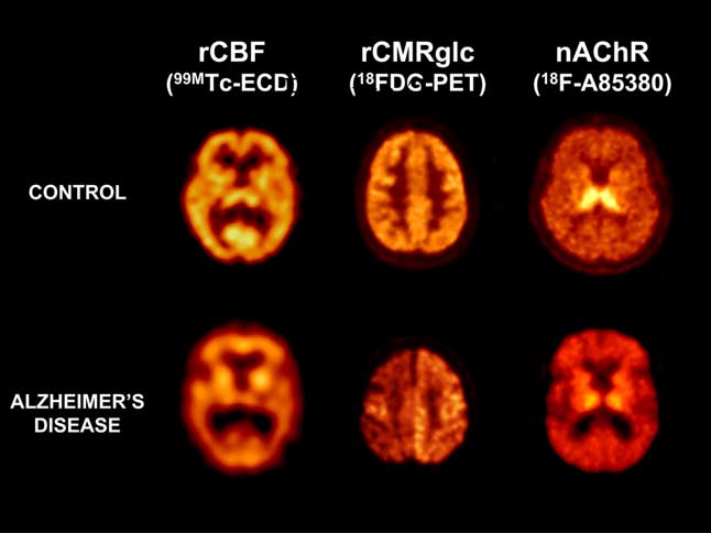
More sensitive functional imaging modalities, such as functionalMR imaging (fMRI), MR Perfusion, MR Spectroscopy (MRS),MR Diffusion weighted imaging, positron emission tomography(PET), and single photon emission computed tomography(SPECT), have the capability to identify subtle pathophysiologicchanges in the brain, before structural changes are pres-ent,29;32;36;39–42 therefore possessing greater potential for accurateand early diagnosis, monitoring disease progression, and bettertreatment follow-up.43 While fMRI detects regional changes in deoxyhemoglobin concentration reflecting focal brain activity in response to avariety of stimuli and memory tasks,44 MR Perfusion has beenshown to have about 85–95% sensitivity for patients with mild ormoderate AD with a range of 88–95% specificity.45;46 Estimation Representative SPECT regional cerebral blood flow (rCBF, left), PET of regional metabolite levels of N-acetylaspartate (NAA), gluta- regional metabolic rates of glucose (rCMRglc, center), and PET nicotinic mine and glutamate, c-aminobutyric acid, myo-inositol, glycine, receptor (nAChR, right) transaxial images in control subjects (top row) and choline, creatine and phosphocreatine, lipids, and lactate in brain Alzheimer’s patients (bottom row).
ª 2004 Elsevier Ltd. All rights reserved.
Journal of Clinical Neuroscience (2005) 12(3), 221–230 described in patients with AD and not in age-matched control subjects nor in patients with other forms of dementia.60;61 Though Extracellular amyloid plaques are the hallmark brain lesions of still not totally accepted as part of the AD patient diagnostic work- sporadic Alzheimer's disease. These microscopic Ab aggregates91 up,62 there is mounting evidence suggesting that incorporation of are well beyond the resolution of the usual neuroimaging tech- FDG–PET into the diagnostic work-up of patients with early niques used for the evaluation of patients with AD. Furthermore, symptoms of cognitive decline might improve diagnostic and current techniques focus on non-specific features derived mainly prognostic accuracy, thereby reducing both disease and treatment- from neuronal loss and atrophy, which are late features in the related morbidity of patients with dementia.63 After examining progression of the disease, and are secondary to the basic func- 129 cognitively impaired patients, the overall sensitivity for tional alteration. However, the distribution and density of both detecting temporoparietal hypometabolism by PET in patients diffuse and Ab plaques at the light microscopic level have not with probable AD was 94%.60 Significantly lower temporoparietal been consistently shown to correlate with the presence or severity metabolic activity was found in asymptomatic subjects with the of dementia.92;93 The best correlation occurs with soluble levels of apolipoprotein e4 allele than in those without the allele.64;65 In a Ab, measured biochemically.91 Soluble Ab is in equilibrium with multicenter study the prognostic value of FDG–PET showed a insoluble Ab in the plaques. Since Ab is at the centre of patho- high degree of sensitivity (93%) and moderate specificity (73%) genesis, many efforts are now focused in developing a radiotracer for prediction of progressive dementia.66 Posterior cingulate and temporoparietal hypometabolism was observed in MCI patients For a radioligand to be useful as a neuroimaging probe for Ab, when compared to controls. Progression of some of these patients a number of key general properties must be present (Table 4).
to probable AD showed an additional bilateral hypometabolism in These Ab probes must be small lipophilic molecules that cross the prefrontal areas, with further reductions in the posterior cingulate blood–brain barrier (BBB) and bind to Ab in a specific and and parietal cortex, while no such changes were observed in the selective fashion. Several such molecules, modified to enhance BBB permeability, show promise as PET or SPECT ligands.
SPECT studies evaluating regional cerebral blood flow (rCBF) Several compounds have been evaluated as potential Ab probes: have shown a similar pattern as the one described for PET–FDG derivatives of histopathological dyes such as Congo red and studies, with relative rCBF paucity in the temporoparietal re- Chrysamine-G, as well as self-associating Ab amyloid fragments gions59;68 (Fig. 2). It has also been used for the differential diag- nosis of dementia.68–72 In a study with histologic confirmation in70 patients with dementia and 85 control subjects,73 a positiveSPECT scan increased the pre-test probability from 84% to 92% in patients with a clinical diagnosis of “probable AD” and from It has been known since the 1930s that Ab amyloid plaques 67% to 84% in patients with “possible AD.” A negative SPECT present in postmortem AD brain tissue can be stained for histo- scan decreased the probability from 84% to 70% in patients with logical examination with Congo red or Chrysamine-G. Klunk and “probable AD” and from 67% to 52% in patients with “possible colleagues95–97 have developed numerous Congo red derivatives AD.” In a prospective study with histologic confirmation of over for potential use as in vivo Ab amyloid probes, but as relatively 200 dementia cases and 119 control cases,74 SPECT rCBF eval- large and acidic compounds, their ability to cross the blood–brain uation allowed differentiation of patients with AD from control barrier was found to be marginal at best. Recently they have de- subjects with high sensitivity and specificity (89% and 80%, scribed an 11C-labelled methoxy derivative of Congo red [1,4- respectively). On the other hand, patients with MCI followed up bis(40-hydroxystyryl)-2-methoxybenzene] or methoxy-X04 that for three years failed to show a correlation between the presence has much more favourable blood–brain barrier penetration and of a SPECT abnormality at baseline examination and subsequent whose fluorescent properties make it suitable for in vivo dem- cognitive decline in MMSE score,75 while another study showed onstration of Ab accumulation in PS1/APP transgenic mice with more prominent rCBF decreases in mesial temporal lobe and multiphoton microscopy, allowing visualization of individual 1 cingulate gyrus in MCI subjects who subsequently converted to lm plaques within 30–60 min. It was concluded that 11C-labelled AD over a period of 1–2 years.76 Similar reductions were also Methoxy-X04 is a viable candidate as an in vivo Ab amyloid present in asymptomatic subjects with the PS-1 gene mutation, as imaging agent.98 Thioflavin T derivatives have shown even more favourable Ab binding characteristics.99–107 PET studies of 16 AD PET and SPECT can also assess neurotransmitter systems in patients and nine control subjects with 11C N-methyl-[11C]2-(4- vivo. Nicotinic acetylcholine receptors (nAChRs) have been methylaminophenyl)-6-hydroxybenzothiazole implicated in a variety of central processes, such as memory and prominent retention in cortical (such as frontal, parietal, occipital, cognition78;79 (Fig. 2). Abnormally low densities of nAChRs and temporal cortices) and subcortical (striatum) areas known to have been measured in vitro in autopsy brain tissue of AD pa- contain high concentrations of Ab amyloid deposits, while similar tients. There is a great interest to develop radiotracers to image PIB retention was observed in both AD patients and controls in nAChRs non-invasively in order to evaluate receptor impair- areas known for low Ab amyloid deposition (such as white matter ments even at a presymptomatic stage of AD as well as moni- toring drug treatment outcomes.80–84 PET studies revealed a Synthesis and initial characterization of 125I bromostyrylben- reduced uptake and binding of 11C-nicotine in the temporal and zene (BSB) probes were described by Kung and colleagues.109–111 frontal cortices of AD patients.79;85;86 Treatment with cholinergicdrugs in AD patients could lead to recovery of the nAChRs in Ideal characteristics for Ab amyloid radiotracer the brain, as visualized by PET. Tacrine treatment increasedcerebral blood flow, cerebral glucose utilization, and uptake of  Bind selectively and specifically to Ab [11C]nicotine to the brain paralleled by improvement in neuro-  Bind preferably in a reversible fashion psychological performance. Changes in nicotinic receptors and  Readily cross the BBB Minimal systemic metabolism blood flow were observed after three weeks of treatment, while  Provide high signal (Ab) to noise (background, non-specific binding) ratio changes in glucose metabolism were measured after three  Provide quantitative reproducible information about total Ab amyloid burden Journal of Clinical Neuroscience (2005) 12(3), 221–230 ª 2004 Elsevier Ltd. All rights reserved.
Partial list of PET and SPECT Ab amyloid tracers [18F]FDDND, 2-(1-{6-[(2-[18F]-fluoroethyl)(methyl)-amino]-2-naphthalen})- 0.12 Æ 0.02Low affinity site,1.86 Æ 0.22 [18F]FENE, 1-{6-[(2[18F]-fluoroethyl)(methyl)-amino]naphthalen-2-yl]}ethanone High affinity site,0.16 Æ 0.09Low affinity site,71.2 Æ 8.6 [11C], 6-Me-BTA-0 2-(40-aminophenyl)-6-methyl-benzothiazole [11C], 6-Me-BTA-2 2-[40-(dimethylamino)phenyl]-6-methylbenzothiazole [11C], 6-Me-BTA-1 2-(40-methylaminophenyl)-6-methyl-benzothiazole [11C]BTA-1, 2-(40-methylaminophenyl)-benzothiazole [11C]6-OH-BTA-1, 2-(40-methylaminophenyl)-6-hydroxy-benzothiazole [123I/125I]BTA, 2-(30-iodo-40-aminophenyl-6-hydroxy-benzothiazole [125I]TZDM, 2-[40-(dimethylamino)phenyl]-6-iodo-benzothiazole [125I]TZPI, 2-[40-(40000-methyl-piperazin-1-yl)-phenyl]-6-iodobenzothiazole [125I]IMSB, (E,E)-1-iodo-2,5-bis(3-hydrocarbonyl-4-methoxy)-styrylbenzene [125I]ISB, (E,E)-1-iodo-2,5-bis(3-hydrocarbonyl-4-hydroxy)-styrylbenzene [125I]BSB, 1-bromo-2,5-bis-(3-hydrocarbonyl-4-hydroxy)-styrylbenzene [125I]IBOX, 2-(40-dimethylaminophenyl)-6-iodo-benzoxazole [123I/125I]IMPY, 6-iodo-2-(40-dimethylamino)phenyl-imidazo-[1,2-a] pyridine BSB (trans,trans)-1-Bromo-2,5-bis-(3-Hydroxycarbonyl-4-Hydroxy) Styrylbenzene [11C]Methoxy-XO4, 1,4-bis(40-hydroxy-styryl)-2-methoxy-benzene [111In]-DTPA- Ab ð3–40Þ Diethylenetriaminepenta-acetic acid PBN N-tert-butyl-a-phenylnitrore “20,70-dichlorodihydrofluorescein & [18F]BF-108, 3-(2-[18F] fluoroethyl)-ethylamino-6-diethyl-aminoacridine Other BSB isomers were radio-labelled by the same group and slower clearance of [18F]FDDNP than controls in brain areas such their in vitro binding properties to postmortem AD tissues as the hippocampus most affected by plaque deposition. Retention assessed.112–114 All these compounds were found to strongly bind time of [18F]FDDNP in these brain regions was correlated with to Ab amyloid plaques as assessed by fluorescent microscopy, but lower memory performance scores in patients with AD.129 displayed low in vivo brain uptake. Some additional limitations tothe potential use of styrylbenzene derivatives for human in vivo amyloid imaging were described where BSB was shown to notonly bind to Ab amyloid deposits, but also to neurofibrillary Anti-Ab monoclonal antibodies that bind to specific epitopes tangles, neuropil threads, hyperphosphorylated tau protein, and within Ab amyloid fibrils have been developed and used to image Lewy Bodies (synuclein proteins) and related cytoplasmic inclu- amyloid deposits in human brains tissue in vitro.130;131 Because sions that are found in multisystem atrophy.115 Based on these antibodies are poorly delivered into the CNS when administered findings, new PET and SPECT potential compounds are now peripherally, they have usually failed as tracers for in vivo brain being further characterized by this group.116–122 Observations that Ab is a self-aggregating peptide with a very high affinity to itself led to the assessment of radio-labelled modified Ab fragments as in vivo probes132–136, but met with limited success A very lipophilic radiofluorinated 6-dialkylamino-2-naphthyethy- because of poor BBB penetration and rapid degradation.137;138 lidene derivative that presents nanomolar affinity to two distinct Other accessory Ab plaque molecules such as serum amyloid P binding sites on Ab fibrils was developed and characterized by and basic fibroblast growth factor,139 a variety of other small Barrio and colleagues.123–125 2-(1-{6-[(2-[18F]Fluoroethyl) (me- compounds such as analogs of acridine orange,140 and 20,70- thyl) amino]-2naphthyl}ethylidene) malononitrile or [18F]FDDNP dichlorodihydrofluorescein and 10-acetyl-3,7-dihydroxyphenox- is reported to bind both the extracellular Ab plaques and the intra- azine which become fluorescent after oxidation141 have been also cellular NFTs in AD126 while also binding to prion plaques from Creutzfeldt–Jakob Disease.127 [18F]FDDNP was used to obtain thefirst human PET images of Ab in an 82-year-old woman with AD.
Briefly, [18F]FDDNP showed a differential clearance, being slowerfrom areas of plaque deposition, such as the hippocampus, as Neuronal degeneration with impairment in cholinergic transmis- pathologically confirmed later at postmortem examination.128 sion in hippocampal and cortical areas associated with memory and In a follow-up study, both AD patients (n ¼ 9) and control cognition are characteristic of AD. No current therapy has been subjects (n ¼ 7) underwent [18F]FDDNP PET studies.126 The pa- shown to halt or reverse the underlying disease process. Though tients with AD again demonstrated higher accumulation and now approved for AD, the cholinesterase inhibitors tacrine, ª 2004 Elsevier Ltd. All rights reserved.
Journal of Clinical Neuroscience (2005) 12(3), 221–230 Potential applications of Ab amyloid radiotracers homogenized postmortem human brain samples.147 Cherny andcolleagues147 tested the efficacy of CQ in transgenic Tg2576 mice, expressing mutant APP protein and which develop Ab amyloid deposits, and showed a dramatic 49% decrease in brain Ab  Treatment follow-up Elucidate the relationship between symptomatology and Ab amyloid burden deposition after nine weeks of oral treatment. Furthermore, the treated animals showed improvements of other general parameters(body weight, alertness, motor activity, etc.), though they did notlive longer.
donepezil, rivastigmine, and galantamine only provide patients Based on this therapeutic approach, we have been developing a with modest relief to their symptoms.142 Recently, the non-com- method for direct and quantitative in vivo evaluation of amyloid petitive NMDA antagonist memantine has been proposed as a safe burden using the metal binding sites of Ab as targets for in vivo and effective symptomatic treatment of AD patients.143 Other ap- probes. Studies with 123=125I-labelled CQ in mice and human brain proaches to alter the progression of AD involve the use of estrogen, homogenates commenced, with the aim of using 123I-CQ as an antioxidants (alone or in combination with selegiline), or non- steroidal anti-inflammatory drugs. Potentially, the most promisingstrategy would involve retarding, halting, or even reversing the process that leads to the formation of Ab plaques.142;144 Because we are now approaching a point at which several While clinical criteria together with current structural neuroim- pharmacological agents aimed at reducing levels of Ab in the aging techniques (CT or MRI) are sensitive and specific enough brain are being developed and tested, the importance of the in vivo for the diagnosis of AD at the mid or late stages of the disease, the evaluation of the Ab amyloid burden is highlighted (Table 6).
development of a reliable method of assessing Ab amyloid burden Given the evidence that levels of soluble Ab correlate with in vivo may permit early diagnosis at presymptomatic stages, disease severity91;145 and that the Ab amyloid is probably the main more accurate differential diagnosis, while also allowing treat- neurotoxic factor in the development of AD, two basic strategies have been proposed in order to reduce or remove Ab from the The criteria for the diagnosis, management, and early detection brain: immunization146 and breaking the pathway that leads to Ab of dementia62;167;168 published by the American Academy of Neurology Quality Standards Subcommittee supports the use of The first approach, which has been shown to be effective in CT and MRI in the work-up of the patient with dementia, while mice models of AD, relies on either precipitating an active im- recommending further research to determine the utility of other mune response against the Ab,146;148;149 or the passive adminis- neuroimaging modalities such as PET and, to a lesser degree tration of specific anti-Ab antibodies.150;151 SPECT.62 Though FDG PET is mainly used in the differential Based on the role that metal ions may play in the biochemical diagnosis of AD, it is the neuroimaging technique that has been processes associated with Ab deposition and neurotoxicity,152–159 a shown to yield the highest prognostic value for providing a further therapeutic strategy using the metal binding sites of Ab as diagnosis of presymptomatic AD two or more years before the full potential targets has been developed. Briefly, Ab is a metallo- dementia picture is manifested.66;169–171 Given the growing evi- protein, with high in vitro affinity for Cu2þ (highest), Fe3þ and dence, PET will likely come to be at the forefront of the AD Zn2þ (lowest).153;154;160;161 Association of soluble Ab with both neuroimaging tools both as a diagnostic as well as a prognostic Fe3þ and Cu2þ produces H2O2, which is neurotoxic in vitro,155–157 tool, providing new insights into the spatial and temporal pattern while complexing of Ab with redox-inert Zn2þ causes precipita- tion of the soluble metalloprotein complex.158 Elevated levels of Because new treatment strategies to prevent or slow disease Cu2þ, Fe3þ, and Zn2þ are also found in the Ab amyloid deposits of progression through early-intervention are being developed and AD brains, thereby demonstrating in vivo what is observed in implemented, there is an urgent need for early disease recognition, vitro.159 Addition of Zn2þ at a molar excess to Cu2þ- and/or Fe3þ which is reflected in the necessity of developing sensitive and bound Ab prevents the production of H2O2 and the corresponding specific biomarkers, specific for a particular trait underlying the neurotoxicity, possibly by displacement of the bound Cu2þ and/or pathological process, as adjuncts to clinical and neuropsycholog- Fe3þ.157 Assessing the level of oxidative damage in vivo in AD- affected tissue by measuring the levels of 8-OH guanosine reveals But the emphasis should not be limited to the ability of early an inverse relationship between amyloid burden and oxidative diagnosis. With new therapeutic approaches being developed that damage.157 This implies that the presence of Ab amyloid could be either prevent the deposition of Ab or increase its solubilization, a response to increased oxidative damage caused by metal-bound agents that could delay the onset of dementia, the role of soluble Ab, and that Zn2þ binding precipitates Ab amyloid and imaging and quantifying Ab amyloid in vivo is becoming cru- thereby neutralizes the toxic soluble Ab.162;163 Metal-protein cial. The ability to detect preclinical or early stage disease attenuating compounds (MPAC) not only inhibit the in vitro through clinical, laboratory, and neuroimaging tests, combined generation of hydrogen peroxide but also have been shown to with anti-Ab amyloid in the at-risk patient, or the patient with reverse the precipitation of Ab in vitro and in post mortem human MCI, may prevent or delay functional and irreversible cognitive brain specimens.164 Thus, this second strategy using MPACs could losses, allowing at the same time to customize and monitor operate in a twofold fashion: it would reduce Ab amyloid burden by a direct solubilization effect and also reduce toxic oxidative One day in the near future we may be able to say: Thou by thy dial’s shady stealth mayst know/Time’s thievish progress to Clioquinol (CQ), ‘5-chloro-7-iodo-8-hydroxyquinoline’ is a eternity./Look, what thy memory can not contain/Commit to these hydrophobic quinoline Zn2þ and Cu2þ chelator that freely crosses waste blanks, and thou shalt find/Those children nursed, deliver’d the blood–brain barrier.165 CQ was chosen to be tested as an Ab from thy brain,/To take a new acquaintance of thy mind.* amyloid solubilizing and anti-toxic agent in Phase II clinical tri-als166 after initial studies showed that CQ increased soluble phaseAb by more than 200% in a concentration-dependent fashion in Journal of Clinical Neuroscience (2005) 12(3), 221–230 ª 2004 Elsevier Ltd. All rights reserved.
Jobst KA, Smith AD, Szatmari M et al. Detection in life of confirmedAlzheimer’s idsease using a simple measurement of medial temporal lobe This work is supported in part by grants from the National Health atrophy by computed tomography. Lancet 1992; 340(8829): 1179–1183.
and Medical Research Council of Australia, Prana Biotechnology, Scheltens PH. Structural neuroimaging of Alzheimer’s disease and other dementias. Aging 2001; 13(3): 203–209.
O’Brien JT, Metcalfe S, Swann A et al. Medial temporal lobe width on CTscanning in Alzheimer’s disease; comparison with vascular dementia, depression and dementia with Lewy bodies. Dement Geriatr Cogn Disord2000; 11(2): 114–118.
Khachaturian ZS. Diagnosis of Alzheimer’s disease. Arch Neurol 1985; Juottonen K, Laakso MP, Insausti R et al. Volumes of the entorhinal and perirhinal cortices in Alzheimer’s disease. Neurobiol Aging 1998; 19(1): 15– Cummings JL, Vinters HV, Cole GM, Khachaturian ZS. Alzheimer’s disease: etiologies, pathophysiology, cognitive reserve, and treatment opportunities.
Du AT, Schuff N, Amend D et al. Magnetic resonance imaging of the Neurology 1998; 51(Suppl 1): S2–S17, discussion S65–7.
entorhinal cortex and hippocampus in mild cognitive impairment and Larson EB, Edwards JK, O’Meara E, Nochlin D, Sumi SM. Neuropathologic Alzheimer’s disease. J Neurol Neurosurg Psychiatry 2001; 71(4): 441–447.
diagnostic outcomes from a cohort of outpatients with suspected dementia. J Wolf H, Jelic V, Gertz HJ, Nordberg A, Julin P, Wahlund LO. A critical Gerontol A Biol Sci Med Sci 1996; 51(Suppl 6): M313–M318.
discussion of the role of neuroimaging in mild cognitive impairment. Acta Rasmusson DX, Brandt J, Steele C, Hedreen JC, Troncoso JC, Folstein MF.
Neurol Scand Suppl 2003; 179: 52–76.
Accuracy of clinical diagnosis of Alzheimer disease and clinical features of Xu Y, Jack CRJ, O’Brien PC et al. Usefulness of MRI measures of entorhinal patients with non-Alzheimer disease neuropathology. Alzheimer Dis Assoc cortex versus hippocampus in AD. Neurology 2000; 54(9): 1760–1767.
Barber R, McKeith IG, Ballard C, Gholkar A, O’Brien JT. A comparison of American Psychiatric Association DSM-IV: Diagnostic and Statistical Manual medial and lateral temporal lobe atrophy in dementia with Lewy bodies and (fourth edn). DSM-IV: Diagnostic and Statistical Manual (fourth edn).
Alzheimer’s disease: magnetic resonance imaging volumetric study. Dement American Psychiatric Association, Washington, DC; 1994.
Geriatr Cogn Disord 2001; 12(3): 198–205.
International Classification of Diseases (ICD-10): Classification of Mental and Chetelat G, Baron JC. Early diagnosis of Alzheimer’s disease; contribution of Behavioural Disorders (10th edn). World Health Organization, Geneva, structural neuroimaging. Neuroimage 2003; 18(2): 525–541.
Scheltens P, Fox N, Barkhof F, De Carli C. Structural magnetic resonance McKhann G, Drachman D, Folstein M, Katzman R, Price D, Stadlan EM.
imaging in the practical assessment of dementia: beyond exclusion. Lancet Clinical diagnosis of Alzheimer’s disease: report of the NINCDS-ADRDA Work Group under the auspices of Department of Health and Human Services Killiany RJ, Gomez-Isla T, Moss M et al. Use of structural magnetic resonance Task Force on Alzheimer’s Disease. Neurology 1984; 34: 939–944.
imaging to predict who will get Alzheimer’s disease. Ann Neurol 2000; 47(4): Petersen RC. Mild cognitive impairment: transition between aging and Alzheimer’s disease. Neurologia 2000; 15(3): 93–101.
Killiany RJ, Hyman BT, Gomez-Isla T et al. MRI measures of entorhinal cortex Petersen RC, Smith GE, Ivnik RJ et al. Apolipoprotein E status as a predictor vs hippocampus in preclinical AD. Neurology 2002; 58(8): 1188–1196.
of the development of Alzheimer’s disease in memory-impaired individuals.
Petrella JR, Coleman RE, Doraiswamy PM. Neuroimaging and early diagnosis of Alzheimer disease: a look to the future. Radiology 2003; 226(2): 315–336.
Petersen RC, Smith GE, Waring SC, Ivnik RJ, Tangalos EG, Kokmen E. Mild de Leon MJ, Convit A, DeSanti S et al. Contribution of structural cognitive impairment: clinical characterization and outcome. Arch Neurol neuroimaging to the early diagnosis of Alzheimer’s disease. Int Psychogeriatr 1997; 9(Suppl 1): 183–190, discussion 247–52.
Miklossy J, Taddei K, Suva D et al. Two novel presenilin-1 mutations (Y256S De Toledo-Morrell L, Goncharova I, Dickerson B, Wilson RS, Bennett DA.
and Q222H) are associated with early-onset Alzheimer’s disease. Neurobiol From healthy aging to early Alzheimer’s disease: in vivo detection of entorhinal cortex atrophy. Ann N Y Acad Sci 2000; 911: 240–253.
Rocchi A, Pellegrini S, Siciliano G, Murri L. Causative and susceptibility Bobinski M, de Leon MJ, Convit A et al. MRI of entorhinal cortex in mild genes for Alzheimer’s disease: a review. Brain Res Bull 2003; 61(1): 1–24.
Alzheimer’s disease. Lancet 1999; 353(9146): 38–40.
Mayeux R, Saunders AM, Shea S et al. Utility of the apolipoprotein E Dickerson BC, Goncharova I, Sullivan MP et al. MRI-derived entorhinal and genotype in the diagnosis of Alzheimer’s disease: Alzheimer’s Disease hippocampal atrophy in incipient and very mild Alzheimer’s disease.
Centers Consortium on apolipoprotein E and Alzheimer’s disease. N Engl J Neurobiol Aging 2001; 22(5): 747–754.
Silverman DH, Phelps ME. Application of positron emission tomography for Corder EH, Saunders AM, Strittmatter WJ et al. Gene dose of apolipoprotein E evaluation of metabolism and blood flow in human brain: normal type 4 allele and the risk of Alzheimer’s disease in late onset families. Science development, aging, dementia, and stroke. Mol Genet Metab 2001; 74(1–2): O’Brien J, Ames D, Burns A (eds) Dementia. second edn. Arnold, London Courtney SM, Ungerleider LG, Keil K, Haxby JV. Transient and sustained activity in a distributed neural system for human working memory. Nature Jellinger K, Maurer K, Riederer P, Beckmann H (eds) Alzheimer Disease: Epidemiology, Neuropathology and Clinics Morphology of Alzheimer Disease Harris GJ, Lewis RF, Satlin A et al. Dynamic susceptibility contrast MR and Related Disorders. Springer, New York, NY 1990; 61–77.
imaging of regional cerebral blood volume in Alzheimer disease: a promising Selkoe DJ. Alzheimer’s disease: genotypes, phenotypes, and treatments.
alternative to nuclear medicine. AJNR Am J Neuroradiol 1998; 19(9): 1727– Michaelis ML, Dobrowsky RT, Li G. Tau neurofibrillary pathology and Bozzao A, Floris R, Baviera ME, Apruzzese A, Simonetti G. Diffusion and microtubule stability. J Mol Neurosci 2002; 19(3): 289–293.
perfusion MR imaging in cases of Alzheimer’s disease: correlations with Jellinger KA, Bancher C. Neuropathology of Alzheimer’s disease: a critical cortical atrophy and lesion load. AJNR Am J Neuroradiol 2001; 22(6): 1030– update. J Neural Transm Suppl 1998; 54: 77–95.
Perl DP. Neuropathology of Alzheimer’s disease and related disorders. Neurol Doraiswamy PM, Chen JG, Charles HC. Brain magnetic resonance spectroscopy: role in assessing outcomes in Alzheimer’s disease. CNS Drugs Geula C. In: Scinto LFM, Daffner KR (eds) Pathological Diagnosis of Alzheimer’s Disease The Early Diagnosis of Alzheimer’s Disease. Humana, Jessen F, Block W, Traber F et al. Proton MR spectroscopy detects a relative decrease of N-acetylaspartate in the medial temporal lobe of patients with AD.
Martins RN, Robinson PJ, Chleboun JO, Beyreuther K, Masters CL. The molecular pathology of amyloid deposition in Alzheimer’s disease. Mol Longo R, Giorgini A, Magnaldi S, Pascazio L, Ricci C. Alzheimer’s disease Neurobiol 1991; 5(2–4): 389–398.
histologically proven studied by MRI and MRS: two cases. Magn Reson Beyreuther K, Masters CL. Amyloid precursor protein (APP) and beta A4 amyloid in the etiology of Alzheimer’s disease: precursor–product Miller EK, Li L, Desimone R. Activity of neurons in anterior inferior relationships in the derangement of neuronal function. Brain Pathol 1991; 1(4): temporal cortex during a short-term memory task. J Neurosci 1993; 13(4): Masters CL, Beyreuther K, Henryk M. Wisniewski and the amyloid theory of Frederick BB, Satlin A, Yurgelun-Todd DA, Renshaw PF. In vivo proton Alzheimer’s disease. J Alzheimers Dis 2001; 3(1): 83–86.
magnetic resonance spectroscopy of Alzheimer’s disease in the parietal and Selkoe DJ. In: Scinto LFM, Daffner KRE (eds) The Early Diagnosis of temporal lobes. Biol Psychiatry 1997; 42: 147–150.
Alzheimer’s Disease The Pathophysiology of Alzheimer’s Disease. Humana, Klunk WE, Panchalingam K, Moossy J, McClure RJ, Pettegrew JW. Acetyl-L- aspartate and other amino acid metabolites in Alzheimer’s disease brain: a ª 2004 Elsevier Ltd. All rights reserved.
Journal of Clinical Neuroscience (2005) 12(3), 221–230 preliminary proton nuclear magnetic resonance study. Neurology 1992; 42(8): Villemagne VL, Musachio JL, Scheffel U et al. Nicotine and related compounds as PET and SPECT ligands. In: Neuronal Nicotinic Receptors: Mohanakrishnan P, Fowler AH, Vonsattel JP et al. An in vitro 1H nuclear Pharmacology and Therapeutic Opportunities. Wiley, New York 1998; magnetic resonance study of the temporoparietal cortex of Alzheimer brains.
Exp Brain Res 1995; 102(3): 503–510.
Nordberg A, Hartvig P, Lilja A et al. Nicotine receptors in the brain of patients Satlin A, Bodick N, Offen WW, Renshaw PF. Brain proton magnetic with Alzheimer’s disease. Studies with 11C-nicotine and positron emission resonance spectroscopy (H-MRS) in Alzheimer’s disease: changes after tomography. Acta Radiol Suppl 1991; 376: 165–166.
treatment with xanomeline, an M1 selective cholinergic agonist. Am J Volkow ND, Ding YS, Fowler JS, Gatley SJ. Imaging brain cholinergic Psychiatry 1997; 154(10): 1459–1461.
activity with positron emission tomography: its role in the evaluation of Waldman AD, McConnell JR, Rai GS, Chaudry M, Grant DS, Martin PA.
cholinergic treatments in Alzheimer’s dementia. Biol Psychiatry 2001; 49(3): Brain MR spectroscopy at 1.0 tesla. Br J Radiol 1997; 70(836): 867.
Kantarci K, Jack CR, Xu YC et al. Mild cognitive impairment and Alzheimer Sihver W, Langstrom B, Nordberg A. Ligands for in vivo imaging of nicotinic disease: regional diffusivity of water. Radiology 2001; 219(1): 101–107.
receptor subtypes in Alzheimer brain. Acta Neurol Scand Suppl 2000; 176: Rose SE, Chen F, Chalk JB et al. Loss of connectivity in Alzheimer’s disease: an evaluation of white matter tract integrity with colour coded MR diffusion tensor Villemagne VL, Horti A, Scheffel U et al. Imaging nicotinic acetylcholine imaging. J Neurol Neurosurg Psychiatry 2000; 69(4): 528–530.
receptors with PET and [F-18]-FPH, a [F-18]-labeled analog of epibatidine. J Phelps ME. PET: the merging of biology and imaging into molecular imaging.
Musachio JL, Villemagne VL, Scheffel U et al. [125/123I]IPH: a Camargo EE. Brain SPECT in neurology and psychiatry. J Nucl Med 2001; radioiodinated analog of epibatidine for in vivo studies of nicotinic acetylcholine receptors. Synapse 1997; 26(4): 392–399.
Salmon E, Sadzot B, Maquet P et al. Differential diagnosis of Alzheimer’s Musachio JL, Villemagne VL, Scheffel UA et al. Synthesis of an I-123 analog disease with PET. J Nucl Med 1994; 35(3): 391–398.
of A-85380 and preliminary SPECT imaging of nicotinic receptors in baboon.
Devanand DP, Jacobs DM, Tang MX et al. The course of psychopathologic Nucl Med Biol 1999; 26(2): 201–207.
features in mild to moderate Alzheimer disease. Arch Gen Psychiatry 1997; Nordberg A. In vivo detection of neurotransmitter changes in Alzheimer’s disease. Ann N Y Acad Sc 1993; 695: 27–33.
Knopman DS, DeKosky ST, Cummings JL et al. Practice parameter: diagnosis Nordberg A. Clinical studies in Alzheimer patients with positron emission of dementia (an evidence based review). Report of the Quality Standards tomography. Behav Brain Res 1993; 57(2): 215–224.
Subcommittee of the American Academy of Neurology. Neurology 2001; Nordberg A, Amberla K, Shigeta M et al. Long-term tacrine treatment in three mild Alzheimer patients: effects on nicotinic receptors, cerebral blood flow, Silverman DH, Cummings JL, Small G et al. Added clinical benefit of glucose metabolism, EEG, and cognitive abilities. Alzheimer Dis Assoc incorporating 2-deoxy-2-[18F]fluoro-D-glucose with positron emission tomography into the clinical evaluation of patients with cognitive impairment.
Nordberg A, Lundqvist H, Hartvig P et al. Imaging of nicotinic and muscarinic Mol Imaging Biol 2002; 4(4): 283–289.
receptors in Alzheimer’s disease: effect of tacrine treatment. Dement Geriatr Kennedy AM, Frackowiak RS, Newman SK et al. Deficits in cerebral glucose metabolism demonstrated by positron emission tomography in individuals at Nordberg A. Effect of long-term treatment with tacrine (THA) in Alzheimer’s risk of familial Alzheimer’s disease. Neurosci Lett 1995; 186(1): disease as visualized by PET. Acta Neurol Scand Suppl 1993; 149: 62–65.
Nordberg A, Lilja A, Lundqvist H et al. Tacrine restores cholinergic nicotinic Small GW, Mazziotta JC, Collins MT et al. Apolipoprotein E type 4 allele and receptors and glucose metabolism in Alzheimer patients as visualized by cerebral glucose metabolism in relatives at risk for familial Alzheimer disease.
positron emission tomography. Neurobiol Aging 1992; 13(6): 747–758.
McLean CA, Cherny RA, Fraser FW et al. Soluble pool of Ab amyloid as a Silverman DH, Small GW, Chang CY et al. Positron emission tomography in determinant of severity of neurodegeneration in Alzheimer’s disease. Ann evaluation of dementia: regional brain metabolism and long-term outcome.
Mega MS, Chu T, Mazziotta JC et al. Mapping biochemistry to metabolism: Drzezga A, Lautenschlager N, Siebner H et al. Cerebral metabolic changes FDG–PET and amyloid burden in Alzheimer’s disease. Neuroreport 1999; accompanying conversion of mild cognitive impairment into Alzheimer’s disease: a PET follow-up study. Eur J Nucl Med Mol Imaging 2003; 30(8): Greenberg SM, Rebeck GW, Vonsattel JP, Gomez-Isla T, Hyman BT.
Apolipoprotein E epsilon 4 and cerebral hemorrhage associated with amyloid Talbot PR, Lloyd JJ, Snowden JS, Neary D, Testa HJ. A clinical role for angiopathy. Ann Neurol 1995; 38(2): 254–259.
99mTc- HMPAO SPECT in the investigation of dementia? J Neurol McLean CA, Beyreuther K, Masters CL. Amyloid Abeta levels in Alzheimer’s Neurosurg Psychiatry 1998; 64: 306–313.
disease – a diagnostic tool and the key to understanding the natural history of Charpentier P, Lavenu I, Defebvre L et al. Alzheimer’s disease and Abeta? J Alzheimers Dis 2001; 3(3): 305–312.
frontotemporal dementia are differentiated by discriminant analysis applied to Link CD, Johnson CJ, Fonte V et al. Visualization of fibrillar amyloid deposits Tc HmPAO SPECT data. J Neurol Neurosurg Psychiatry 2000; 69(5): 661– in living, transgenic Caenorhabditis elegans animals using the sensitive amyloid dye, X-34. Neurobiol Aging 2001; 22(2): 217–226.
Sjogren M, Gustafson L, Wikkelso C, Wallin A. Frontotemporal dementia can Klunk WE, Debnath ML, Pettegrew JW. Development of small molecule be distinguished from Alzheimer’s disease and subcortical white matter probes for the beta-amyloid protein of Alzheimer’s disease. Neurobiol Aging dementia by an anterior-to-posterior rCBFSPET ratio. Dement Geriatr Cogn Bacskai BJ, Klunk WE, Mathis CA, Hyman BT. Imaging amyloid-beta Ishii K, Yamaji S, Kitagaki H, Imamura T, Hirono N, Mori E. Regional deposits in vivo. J Cereb Blood Flow Metab 2002; 22(9): 1035–1041.
cerebral blood flow difference between dementia with Lewy bodies and AD.
Klunk WE, Bacskai BJ, Mathis CA et al. Imaging Ab plaques in living transgenic mice with multiphoton microscopy and methoxy-X04, a Lobotesis K, Fenwick JD, Phipps A et al. Occipital hypoperfusion on systemically administered Congo red derivative. J Neuropath Exp Neurol SPECT in dementia with Lewy bodies but not AD. Neurology 2001; 56(5): Klunk WE, Wang Y, Huang GF, Debnath ML, Holt DP, Mathis CA.
Jagust W, Thisted R, Devous MDS et al. SPECT perfusion imaging in the Uncharged thioflavin-T derivatives bind to amyloid-beta protein with high diagnosis of Alzheimer’s disease: a clinicalpathologic study. Neurology 2001; affinity and readily enter the brain. Life Sci 2001; 69(13): 1471–1484.
Klunk WE, Wang Y, Huang GF et al. The binding of 2-(4’- Jobst KA, Barnetson LP, Shepstone BJ. Accurate prediction of histologically methylaminophenyl)benzothiazole to postmortem brain homogenates is confirmed Alzheimer’s disease and the differential diagnosis of dementia: the dominated by the amyloid component. J Neurosci 2003; 23(6): 2086–2092.
use of NINCDS-ADRDA and DSM-III-R criteria, SPECT, X-ray CT, and Apo Bacskai BJ, Hickey GA, Skoch J et al. Four-dimensional multiphoton E4 in medial temporal lobe dementias: Oxford Project to Investigate Memory imaging of brain entry, amyloid binding, and clearance of an amyloid-beta and Aging. Int Psychogeriatr 1998; 10: 271–302.
ligand in transgenic mice. Proc Natl Acad Sci USA 2003; 100(21): 12462– McKelvey R, Bergman H, Stern J, Rush C, Zahirney G, Chertkow H. Lack of prognostic significance of SPECT abnormalities in non-demented elderly Mathis CA, Bacskai BJ, Kajdasz ST et al. A lipophilic thioflavin-T derivative subjects with memory loss. Can J Neurol Sci 1999; 26: 23–28.
for positron emission tomography (PET) imaging of amyloid in brain. Bioorg Johnson KA, Jones K, Holman BL et al. Preclinical prediction of Alzheimer’s Med Chem Lett 2002; 12(3): 295–298.
disease using SPECT. Neurology 1998; 50(6): 1563–1571.
Mathis CA, Wang Y, Holt DP, Huang GF, Debnath ML, Klunk WE. Synthesis Johnson KA, Lopera F, Jones K et al. Presenilin-1-associated abnormalities in and evaluation of 11C-labeled 6-substituted 2-arylbenzothiazoles as amyloid regional cerebral perfusion. Neurology 2001; 56(11): 1545–1551.
imaging agents. J Med Chem 2003; 46(13): 2740–2754.
Journal of Clinical Neuroscience (2005) 12(3), 221–230 ª 2004 Elsevier Ltd. All rights reserved.
Wang Y, Klunk WE, Huang GF, Debnath ML, Holt DP, Mathis CA. Synthesis Small GW, Agdeppa ED, Kepe V, Satyamurthy N, Huang SC, Barrio JR. In and evaluation of 2-(30-iodo-40-aminophenyl)-6-hydroxybenzothiazole for in vivo brain imaging of tangle burden in humans. J Mol Neurosci 2002; 19(3): vivo quantitation of amyloid deposits in Alzheimer’s disease. J Mol Neurosci Majocha RE, Reno JM, Friedland RP, Van Haight C, Lyle LR, Marotta CA.
Wang Y, Mathis CA, Huang GF et al. Effects of lipophilicity on the affinity Development of a monoclonal antibody specific for b/A4 amyloid in and nonspecific binding of iodinated benzothiazole derivatives. J Mol Alzheimer’s disease brain for application to in vivo imaging of amyloid angiopathy. J Nucl Med 1992; 33(12): 2184–2189.
Helmuth L. Long-awaited technique spots Alzheimer’s toxin. Science 2002; Walker LC, Price DL, Voytko ML, Schenk DB. Labelling of cerebral amyloid in vivo with a monoclonal antibody. J Neuropathol Exp Neurol 1994; 53(4): Mathis CA, Holt DP, Wang Y, Huang GF, Debnath ML, Klunk WE.
Lipophilic 11C-labelled thioflavin-T analogues for imaging amyloid plaques Lee VM. Related amyloid binding ligands as Alzheimer’s disease therapies.
in Alzheimer’s disease. J Label Cpd Radiopharm 2001; 44(Suppl 1): S26– Neurobiol Aging 2002; 23(6): 1039–1042.
Marshall JR, Stimson ER, Ghilardi JR, Vinters HV, Mantyh PW, Maggio JE.
Klunk WE, Engler H, Nordberg A et al. Imaging brain amyloid in Alzheimer’s Noninvasive imaging of peripherally injected Alzheimer’s disease type disease with Pittsburgh compound-B. Ann Neurol 2004; 55: 306–319.
synthetic A beta amyloid in vivo. Bioconjug Chem 2002; 13(2): Zhuang ZP, Kung MP, Hou C et al. Radioiodinated styrylbenzenes and thioflavins as probes for amyloid aggregates. J Med Chem 2001; 44(12): Maggio JE, Stimson ER, Ghilardi JR et al. Reversible in vitro growth of Alzheimer disease beta-amyloid plaques by deposition of Kung HF, Lee CW, Zhuang ZP, Kung MP, Hou C, Plossl K. Novel stilbenes as labeled amyloid protein. Proc Natl Acad Sci USA 1992; 89(12): probes for amyloid plaques. J Am Chem Soc 2001; 123(50): 12740–12741.
Skovronsky DM, Zhang B, Kung MP, Kung HF, Trojanowski JQ, Lee VM. In Friedland RP, Shi J, Lamanna JC, Smith MA, Perry G. Prospects for vivo detection of amyloid plaques in a mouse model of Alzheimer’s disease.
noninvasive imaging of brain amyloid beta in Alzheimer’s disease. Ann N Y Proc Natl Acad Sci USA 2000; 97(13): 7609–7614.
Kung MP, Hou C, Zhuang ZP et al. Radioiodinated styrylbenzene derivatives Ghilardi JR, Catton M, Stimson ER et al. Intra-arterial infusion of [125I]A beta as potential SPECT imaging agents for amyloid plaque detection in 1–40 labels amyloid deposits in the aged primate brain in vivo. Neuroreport Alzheimer’s disease. J Mol Neurosci 2002; 19(1–2): 7–10.
Zhuang ZP, Kung MP, Hou C et al. IBOX(2-(40-dimethylaminophenyl)-6- Kurihara A, Pardridge WM. Abeta(1–40) peptide radiopharmaceuticals for iodobenzoxazole): a ligand for imaging amyloid plaques in the brain. Nucl brain amyloid imaging: (111)In chelation, conjugation to poly(ethylene glycol)-biotin linkers, and autoradiography with Alzheimer’s disease brain Lee CW, Zhuang ZP, Kung MP et al. Isomerization of (Z,Z) to (E,E)1-bromo- sections. Bioconjug Chem 2000; 11(3): 380–386.
2, 5-bis-(3-hydroxycarbonyl-4-hydroxy)styrylbenzene in strong base: probes Saito Y, Buciak J, Yang J, Pardridge WM. Vector-mediated delivery of 125I- for amyloid plaques in the brain. J Med Chem 2001; 44(14): 2270–2275.
labeled beta-amyloid peptide A beta 1–40 through the blood–brain barrier and Schmidt ML, Schuck T, Sheridan S et al. The fluorescent Congo red derivative binding to Alzheimer disease amyloid of the A beta 1–40/vector complex. Proc (trans,trans)-1-bromo-2, 5-bis-(3-hydroxycarbonyl-4-hydroxy)styrylbenzene Natl Acad Sci USA 1995; 92(22): 10227–10231.
(BSB) labels diverse b-pleated sheet structures in postmortem human Shi J, Perry G, Berridge MS et al. Labeling of cerebral amyloid beta deposits neurodegenerative disease brains. Am J Pathol 2001; 159(3): 937–943.
in vivo using intranasal basic fibroblast growth factor and serum amyloid P Zhuang ZP, Kung MP, Wilson A et al. Structure–activity relationship of component in mice. J Nucl Med 2002; 43(8): 1044–1051.
imidazo[1, 2-a]pyridines as ligands for detecting beta-amyloid plaques in the Shimadzu H, Suemoto T, Suzuki M et al. A novel probe for imaging amyloid- brain. J Med Chem 2003; 46(2): 237–243.
b: synthesis of F-18 labelled BF-108, an Acridine orange analog. J Label Kung MP, Hou C, Zhuang ZP et al. IMPY: an improved thioflavin-T Compd Radiopharm 2003; 46: 765–772.
derivative for in vivo labeling of beta-amyloid plaques. Brain Res Bull 2002; McLellan ME, Kajdasz ST, Hyman BT, Bacskai BJ. In vivo imaging of reactive oxygen species specifically associated with thioflavine S-positive Ono M, Kung MP, Hou C, Kung HF. Benzofuran derivatives as Abeta- amyloid plaques by multiphoton microscopy. J Neurosci 2003; 23(6): 2212– aggregate-specific imaging agents for Alzheimer’s disease. Nucl Med Biol Emilien G, Beyreuther K, Masters CL, Maloteaux JM. Prospects for Ono M, Wilson A, Nobrega J et al. 11C-labeled stilbene derivatives as Abeta- pharmacological intervention in Alzheimer disease. Arch Neurol 2000; 57(4): aggregate-specific PET imaging agents for Alzheimer’s disease. Nucl Med Rogawski MA, Wenk GL. The neuropharmacological basis for the use of Kung MP, Skovronsky DM, Hou C et al. Detection of amyloid plaques by memantine in the treatment of Alzheimer’s disease. CNS Drug Rev 2003; 9: radioligands for Abeta40 and Abeta42: potential imaging agents in Alzheimer’s patients. J Mol Neurosci 2003; 20(1): 15–24.
LeVine III H. Challenges of targeting Ab fibrillogenesis and other Kung MP, Zhuang ZP, Hou C, Jin LW, Kung HF. Characterization of protein folding disorders. Amyloid: J Protein Fold Disord 2003; 10: radioiodinated ligand binding to amyloid beta plaques.; J Mol Neurosci 2003; Wang J, Dickson DW, Trojanowski JQ, Lee VM. The levels of soluble versus Lee CW, Kung MP, Hou C, Kung HF. Dimethylamino-fluorenes: ligands for insoluble brain Ab distinguish Alzheimer’s disease from normal and detecting beta-amyloid plaques in the brain. Nucl Med Biol 2003; 30(6): 573– pathologic aging. Exp Neurol 1999; 158(2): 328–337.
Schenk D, Barbour R, Dunn W et al. Immunization with amyloid-b attenuates Agdeppa ED, Kepe V, Petri A et al. In vitro detection of (S)-naproxen and Alzheimer-disease-like pathology in the PDAPP mouse. Nature 1999; ibuprofen binding to plaques in the Alzheimer’s brain using the positron emission tomography molecular imaging probe 2-(1-[6-[(2- Cherny RA, Atwood CS, Xilinas ME et al. Treatment with a copper-zinc [(18)F]fluoroethyl)(methyl)amino]-2- naphthyl]ethylidene)malononitrile.
chelator markedly and rapidly inhibits b-amyloid accumulation in Alzheimer’s Neuroscience 2003; 117(3): 723–730.
disease transgenic mice. Neuron 2001; 30(3): 665–676.
Agdeppa ED, Kepe V, Liu J et al. Binding characteristics of radiofluorinated Weiner HL, Lemere CA, Maron R et al. Nasal administration of amyloid-b 6-dialkylamino-2-naphthylethylidene derivatives as positron emission peptide decreases cerebral amyloid burden in a mouse model of Alzheimer’s tomography imaging probes for b-amyloid plaques in Alzheimer’s disease. J disease. Ann Neurol 2000; 48(4): 567–579.
Janus C, Pearson J, McLaurin J et al. Ab-peptide immunization reduces Barrio JR, Huang SC, Cole G et al. PET imaging of tangles and plaques in behavioural impairment and plaques in a model of Alzheimer’s disease. Nature Alzheimer disease with a highly lipophilic probe. J Label Compd Radiopharm Bard F, Cannon C, Barbour R et al. Peripherally administered antibodies Shoghi-Jadid K, Small GW, Agdeppa ED et al. Localisation of neurofibrillary against amyloid b peptide enter the central nervous system and reduce tangles and b-amyloid plaques in the brains of living patients with Alzheimer’s pathology in a mouse model of Alzheimer disease. Nat Med 2000; 6(8): 916– disease. Am J Ger Psychiatry 2002; 10(1): 24–35.
Bresjanac M, Smid LM, Vovko TD, Petric A, Barrio JR, Popovic M.
DeMattos RB, Bales KR, Cummins DJ, Dodart JC, Paul SM, Holtzman DM.
Molecular-imaging probe 2-(1-[6-[(2-fluoroethyl)(methyl) amino]-2- Peripheral anti-Ab antibody alters CNS and plasma Ab clearance and naphthyl]ethylidene) malononitrile labels prion plaques in vitro. J Neurosci decreases brain Ab burden in a mouse model of Alzheimer’s disease. Proc Natl Acad Sci USA 2001; 98(15): 8850–8855.
Agdeppa ED, Kepe V, Shoghi-Jadid K et al. In vivo and in vitro labeling of Cherny RA, Legg JT, McLean CA et al. Aqueous dissolution of Alzheimer’s plaques and tangles in the brain of an Alzheimer’s disease patient: a case disease Ab amyloid deposits by biometal depletion. J Biol Chem 1999; study. J Nucl Med 2001; 42(Suppl 1): 65P.
ª 2004 Elsevier Ltd. All rights reserved.
Journal of Clinical Neuroscience (2005) 12(3), 221–230 Atwood CS, Moir RD, Huang X et al. Dramatic aggregation of Alzheimer Ab age-related cataracts. Novartis Found Symp 2001; 235: 26–38, discussion 38– by Cu(II) is induced by conditions representing physiological acidosis. J Biol Bush AI. Metal complexing agents as therapies for Alzheimer’s disease.
Atwood CS, Huang X, Khatri A et al. Copper catalyzed oxidation of Neurobiol Aging 2002; 23(6): 1031–1038.
Alzheimer Ab. Cell Mol Biol 2000; 46(4): 777–783.
Padmanabhan G, et al. Clioquinol. In: Klauss E, Florey E (eds.).
Huang X, Atwood CS, Hartshorn MA et al. The Ab peptide of Alzheimer’s Analytical Profiles of Drug Substances. Academic Press, New York; 1989: disease directly produces hydrogen peroxide through metal ion reduction.
Biochemistry 1999; 38(24): 7609–7616.
Ritchie CW, Bush AI, Mackinnon A et al. Metal-protein attenuation with Huang X, Cuajungco MP, Atwood CS et al. Cu(II) potentiation of Alzheimer iodochlorhydroxyquin (clioquinol) targeting a amyloid deposition and toxicity Ab neurotoxicity. Correlation with cell-free hydrogen peroxide production and in Alzheimer disease: a pilot phase 2 clinical trial. Arch Neurol 2003; 60: metal reduction. J Biol Chem 1999; 274(52): 37111–37116.
Cuajungco MP, Goldstein LE, Nunomura A et al. Evidence that the b-amyloid Doody RS, Stevens JC, Beck C et al. Practice parameter: management of plaques of Alzheimer’s disease represent the redox-silencing and entombment dementia (an evidence-based review) – report of the Quality Standards of Ab by zinc. J Biol Chem 2000; 275(26): 19439–19442.
Subcommittee of the American Academy of Neurology. Neurology 2001; Bush AI, Pettingell WH, Multhaup G et al. Rapid induction of Alzheimer Ab amyloid formation by zinc. Science 1994; 265(5177): 1464–1467.
Petersen RC, Stevens JC, Ganguli M, Tangalos EG, Cummings JL, DeKosky Lovell MA, Robertson JD, Teesdale WJ, Campbell JL, Markesbery WR.
ST. Practice parameter: early detection of dementia: mild cognitive Copper, iron and zinc in Alzheimer’s disease senile plaques. J Neurol Sci impairment (an evidence-based review)-report of the Quality Standards Bush AI, Masters CL, Tanzi RE. Copper, beta-amyloid, and Alzheimer’s of the American Academy of Neurology. Neurology 2001; 56(9): disease: tapping a sensitive connection. Proc Natl Acad Sci USA 2003; Silverman DH, Chang CY, Cummings JL et al. Prognostic value of regional Bush AI. The metallobiology of Alzheimer’s disease. Trends Neurosci 2003; brain metabolism in evaluation of dementia. J Nucl Med 1999; 40(Suppl 1): Curtain CC, Ali F, Volitakis I et al. Alzheimer’s disease amyloid-b binds Chang CY, Silverman DH. Accuracy of early diagnosis and its impact on the copper and zinc to generate an allosterically ordered membrane-penetrating management and course of Alzheimer’s disease. Expert Rev Mol Diagn 2004; structure containing superoxide dismutase-like subunits. J Biol Chem 2001; Silverman DH, Gambhir SS, Huang HW et al. Evaluating early dementia with Bush AI, Goldstein LE. Specific metal-catalysed protein oxidation reactions in and without assessment of regional cerebral metabolism by PET: a comparison chronic degenerative disorders of ageing: focus on Alzheimer’s disease and of predicted costs and benefits. J Nucl Med 2002; 43(2): 253–266.
Journal of Clinical Neuroscience (2005) 12(3), 221–230 ª 2004 Elsevier Ltd. All rights reserved.

Source: https://nit.felk.cvut.cz/~dark/Vyuka/AST2013/2013_05_02_ctvrtek/tesar_vondrackova_-_detekce_alzheimera/podklady/Imaginem%20oblivionis%20-%20the%20prospects%20of%20neuroimaging%20for%20early.pdf

Microsoft word - publications_2010

Department of Clinical Physiology, Nuclear Medicine and PET Publications 2010 Doctoral theses and PhD theses defended during the year of 2010 De Nijs, R. Corrections in clinical Magnetic Resonance Spectroscopy and SPECT: Motion correction in MR spectroscopy, Downscatter correction in SPECT. Defended March 2nd 2010 at Technical University of Denmark, Department of Informatics and Mathematica
kremersurban.com

DEPARTMENT OF HEALTH & HUMAN SERVICES Food and Drug Administration Rockville, MD 20857ANDA 091624 Kremers Urban Pharmaceuticals Inc. U.S. Agent for: Kudco Ireland Limited Attention: Kurt Zimmer RA Manager 1101 C Avenue West Seymour, IN 47274 Dear Sir: This is in reference to your abbreviated new drug application (ANDA) dated July 15, 2009, submitted pursuant to section 505(j) of the Fe

Copyright © 2008-2018 All About Drugs