Cerebral Blood Flow during Anticipation of PublicSpeaking in Social Phobia: A PET Study
Maria Tillfors, Tomas Furmark, Ina Marteinsdottir, and Mats Fredrikson
Background: The aim was to examine the neural corre-
ously been investigated in animal phobics (Drevets et al
lates of anxiety elicited by the anticipation of public
1995; Wik et al 1996) and normal healthy volunteers
speaking in individuals with social phobia.
(Benkelfat et al 1995; Chua et al 1999; Reiman 1997;
Methods: Positron emission tomography and 15O-water
Reiman et al 1989). In anticipation of fear-provoking
was used to measure regional cerebral blood flow in
stimuli, animal phobics exhibited rCBF decreases in mo-
subjects with DSM-IV defined social phobia during anxi-
dality-specific primary sensory areas, while elevated per-
ety anticipation. Heart rate and subjective anxiety were
fusion was not observed (Drevets et al 1995; Wik et al
also recorded. While being scanned, subjects were speak-
1996). In healthy, normal individuals, the anterior cingu-
ing alone either before or after speaking in public. To
late, insula, temporal, prefrontal, and the orbitofrontal
evaluate anticipatory anxiety we compared individuals
cortices together with the thalamus and the cerebellum
speaking alone before they were speaking in front of anaudience with those who did the reverse.
have been associated with an increased perfusion duringanticipatory anxiety (Benkelfat et al 1995; Chua et al
Results: Heart rate and subjective anxiety measures
1999; Reiman 1997; Reiman et al 1989). In contrast with
confirmed anticipatory anxiety in social phobics whoperformed their private speech before their public. This
the animal phobics, rCBF decreases were not associated
was accompanied by enhanced cerebral blood flow in the
with anticipatory anxiety in healthy normals. Neural ac-
right dorsolateral prefrontal cortex, left inferior temporal
tivity assessed with the electroencephalogram (EEG) has
cortex, and in the left amygdaloid-hippocampal region.
also been reported to change in social phobics during
Brain blood flow was lower in the left temporal pole and
anticipation of a public speech (Davidson et al 2000a). bilaterally in the cerebellum in the anticipation group.
These authors observed a right-sided activation in the
Conclusions: Brain regions with altered perfusion pre-
anterior temporal and lateral prefrontal scalp regions. sumably reflect changes in neural activity associated with
Individuals with social phobia worry excessively (e.g.,
worry about anticipated public performance. We speculate
Clark 1997) and because the hallmark of anticipatory
that anticipatory anxiety in social phobics originates in an
anxiety is worry (Barlow et al 1996), it would be of
affect sensitive fear network encompassing the amygdal-
interest to explore the neural correlates of anticipatory
oid-hippocampal region, prefrontal, and temporal areas.
anxiety in social phobia (social anxiety disorder). Thus,
Biol Psychiatry 2002;52:1113–1119 2002 Society of
the aim of the present study was to examine the neural
correlates of anxiety elicited by the anticipation of a publicspeaking task in individuals with social phobia. Recently,
Key Words: Anticipatory anxiety, social phobia, public
we examined brain activity during symptom provocation
speaking, induced anxiety, positron emission tomography,regional cerebral blood flow
in social phobics and healthy individuals while speaking infront of an audience and alone (Tillfors et al 2001). Toevaluate anticipatory anxiety, we compared the social
Introduction
phobics who were speaking alone before they were speak-ing in front of an audience (i.e., the anticipation group)
Anticipatory anxiety is characterized by worry about with those who were speaking alone after they had been
future events (Barlow et al 1996). Regional cerebral
speaking in front of an audience (i.e., the comparison
blood flow (rCBF) during anticipatory anxiety has previ-
During social anxiety provocation, rCBF increased
From the Department of Social Sciences (MT), O
more in the amygdaloid complex in those with social
Department of Psychology (TF, MF), Uppsala University, Uppsala; Depart-
phobia than in healthy nonphobic individuals (Tillfors et al
ment of Neuroscience, Psychiatry (IM), Uppsala University, Uppsala; and theUppsala University PET Center (MF), Uppsala University, Uppsala, Sweden.
2001). Because numerous previous studies (see Davidson
Address reprint requests to Dr. M. Tillfors, Orebro University, Department of
and Irwin 1999 and Davidson et al 2000b for a review)
Social Sciences, SE-70182 Orebro, Sweden.
Received July 25, 2001; revised March 6, 2002; accepted March 15, 2002.
also have identified the amygdaloid complex to play a
crucial role in the processing of fear and anxiety, we
Trait Anxiety Inventory (STAI-T) (Spielberger et al 1983). Four
performed a hypothesis driven search for amygdala acti-
unpaired t tests performed in Statview 4.5 for Macintosh were
vation in addition to a whole-brain search using traditional
used to evaluate possible group differences in trait anxiety. Methods and Materials
Subjective and physiologic measures of fear and anxiety wereanalyzed with four directional unpaired t tests of significance
performed in Statview 4.5 for Macintosh (SAS Institute Inc.,
This study was part of a larger investigation of the functional
Cary, NC). A one-tailed alpha level of 0.05 was applied.
neuroanatomy of social phobia before and after treatment withcognitive-behavioral therapy and citalopram. The methodologyhas been described in detail elsewhere (Furmark et al in press;
Tillfors et al 2001). Briefly, 18 right-handed previously untreatedpatients (10 men and 8 women) with DSM-IV social phobia with
All individuals with social phobia were given topics of a neutral,
a mean age (Ϯ SD) of 35.2 (Ϯ 7.3) years (range 23– 46) who did
nonemotional content—i.e., they spoke about a travel experience
not medicate were recruited. Criteria for exclusion were: previ-
or a vacation. On a random basis, 20 sec extract from each
ous or current organic brain disorders and somatic diseases,
delivered speech was transformed to text and counted. The
current comorbid Axis I disorders, menopause, and pregnancy.
counting words were analyzed with an unpaired t test of
Screening included psychiatric (structured clinical diagnostic
significance performed in Statview 4.5 for Macintosh.
interviews, SCID; First et al 1998) and medical histories as wellas trait measures of anxiety (Furmark et al in press). SCID-interviews were performed by an experienced psychiatrist. Par-
ticipants refrained from tobacco, alcohol and caffeine 12 hoursbefore the PET investigation. The Uppsala University Medical
SCANNING. Investigations were performed on a GEMS
Faculty Ethical Review Board and the Uppsala University
PC2048 –15B scanner (General Electrics, Milwaukee, WI) with a
Isotope Committee approved the study. Informed consent was
10-cm axial field of view (Holte et al 1989), producing 15 slices
obtained after the procedure had been fully explained.
with a 6.5 mm slice spacing and a 6 mm axial/transaxialresolution. A venous catheter was inserted and the subjects werefixated in the scanner using a commercial headholder with fast
hardening foam. The participants were positioned in the scanner
Subjects lay in the scanner with their eyes open during all tasks.
such that the planes were parallel to a line between the anterior
During scanning, subjects were either speaking in the presence of
and posterior commissure where the most basal slice was below
a surrounding audience (6 – 8 silently observing individuals) or
the orbita. First, a 10-min transmission scan using a rotating
alone during 2.5 min. About 20 min before the initial scan,
68germanium pin source was completed. During the end of the
subjects were instructed to prepare a 2.5 min speech about a
transmission scan, each subject was given a saline injection to
travel experience or a vacation. Each task was repeated and
attenuate novelty effects. Before each emission scan, 700 –1300
scanned twice with the order counterbalanced between subjects.
Mbq of 15O-water in 3– 4 mL of water (approximately 15
To evaluate anticipatory anxiety related to social fear we com-
Mbq/kg body weight) was injected with 12-min intervals. The
pared the nine social phobics who were speaking alone before
subjects were told to start speaking immediately following
they were speaking in front of an audience (i.e., the anticipation
injections and data were collected in fifteen 10-sec frames. Each
group) with the nine who were speaking alone after they had
subject had a total of four emission scans during two conditions
been speaking in front of an audience (i.e., the comparison
presented in counterbalanced order between subjects. Finally, an
group). Men and women were equally distributed in the two
additional injection was performed when the scanner-bed was
groups. Scores on Spielberger’s State Anxiety Inventory
automatically moved back and forth between two positions 10
(STAI-S; 20 – 80) (Spielberger et al 1983) and ratings of fear and
cm apart. Data were collected only at these positions and never
distress, ranging from 0 –100, were obtained immediately after
while the scanner was moving. This last interleaved scan was
each scan. During each scan heart rate was recorded with the
performed to obtain a scan with full axial coverage of the brain,
PSYLAB6 integrated system for psychophysiology (http//:www.
which aids in the stereotactical normalization of the PET-images. IMAGE RECONSTRUCTION. Data from the first 70 sec after
arrival of the bolus to the brain were summed. Images were
reconstructed from the summation after correction for attenua-
The trait anxiety questionnaires included the Social Phobia Scale
tion and scatter by using the transmission scan (Bergstro¨m et al
(SPS) (Mattick and Clarke 1998), the Social Interaction Anxiety
1983). The additional interleaved scan was reconstructed to a
Scale (SIAS) (Mattick and Clarke 1998), the Personal Report of
30-slice image set with a 20 cm coverage in axial (superior to
Confidence as a Speaker (PRCS) (Paul 1966) and Spielberger’s
inferior) direction (Bergstro¨m et al 1982). All images were
Figure 1. Mean values and standard deviations for subjective measures of state anxiety and heart rate during speaking alone before (i.e.,the anticipation group) or after (i.e., the comparison group) speaking in public for 18 individuals with social phobia.
reconstructed to a 128 ϫ 128 matrix with a pixel size of 2 mm
ANATOMICAL STANDARDIZATION. Normalization of all
individual CBF images into a standard brain shape (Greitz et al
The two groups did not differ in trait anxiety as they had
1991) was performed automatically by matching the scan with
similar scores on the SPS (t Ͻ 1, df ϭ 16, ns), the SIAS
20-cm axial coverage to an atlas template (Andersson and
(t Ͻ 1, df ϭ 16, ns), the PRCS (t Ͻ 1, df ϭ 16, ns) and the
Thurfjell 1997). The images from the four emission scans were
STAI-T (t Ͻ 1, df ϭ 16, ns).
automatically aligned to the 20-cm scan (Andersson 1995),bringing them into the stereotactic space and correcting for head
movements between scans. The stereotactic space was definedbased on the postmortem slicing of a single subject in which
The anticipation group as compared with the comparison
brain contours, gyri, sulci, central structures, and Brodmann
group displayed higher heart rate (t ϭ 2.5, df ϭ 16, p Ͻ
areas were defined (Greitz et al 1991). The cerebral brain atlas
.01) and state anxiety ratings (STAI-S: t ϭ 1.8, df ϭ 16,
software (Thurfjell et al 1995) allows for identification, in terms
p Ͻ .05), but did not differ in fear (t Ͻ 1, df ϭ 16, ns) or
of anatomy, cytoarchitecture, and Talairach coordinates (Ta-
distress (t Ͻ 1, df ϭ 16, ns) (See Figure 1). BLOOD FLOW RECORDINGS. An ANOVA (Friston et al
1995) with one between group factor (the anticipation group vs. the comparison group) was used. To account for between subject
When counting words it was found that there were no
variance and to evaluate anticipatory anxiety we only used the
significant difference between the anticipation group and
first of the two normalized rCBF images from the speaking alone
the comparison group (t ϭ Ϫ.93; df ϭ 16; ns).
condition to be compatible to a random effects model (Peterssonet al 1999). Global flow was estimated using a predefined mask
outlining the brain, but excluding all voxels which changed as aconsequence of study conditions using F-map masking (Anders-
The state related anxiety reaction associated with antici-
son 1997). PET data were normalized for global flow using linear
pation was accompanied by enhanced rCBF in the right
scaling (Andersson 1997). The contrast generated a t map that
dorsolateral prefrontal and left inferior temporal cortices,
was subsequently converted to a z score map through a proba-
as well as in the left amygdaloid-hippocampal region (x ϭ
bility preserving transformation (Friston et al 1991). The signif-
Ϫ27, y ϭ Ϫ13, z ϭ Ϫ13; maximum z value ϭ 2.58, p ϭ
icance of the z score maps was evaluated at an omnibus level
.005 uncorrected for multiple comparisons) (See Figure 2
using the mean square z score (Worsley et al 1995). Local
changes were evaluated using the spatial extent of connected
Brain blood flow was lowered in the left temporal pole
clusters of voxels with a z score above 2.6 (Friston et al 1994). This test takes into account multiple comparisons and has a
and bilaterally in the cerebellum in the anticipation as
cluster-localizing power (Friston et al 1996). The directed search
compared with the comparison group (See Table 1).
for amygdala activation was performed by comparing the two
Because the design is unbalanced in that individuals in
groups using a z threshold of 2.58 corresponding to an uncor-
the anticipation group always performed their private
rected (one tailed) p of .005.
speech first, we performed a linear trend analysis to
43, z ϭ 32). Thus, task repetition did not influence rCBFin structures associated with anticipatory anxiety except inthe prefrontal cortex where rCBF was attenuated by taskrepetition, but elevated as a function of anticipatoryanxiety. Anticipatory related rCBF-increases were ob-served in the dorsolateral prefrontal cortex while repetitionrelated decreases were located in the prefrontal cortexsuperior to the anticipatory cluster. Because the oppositedirection of change and since the centers of gravity ofthese clusters were separated by 28 mm, it is most likelythat anticipatory anxiety rather than task repetition ac-counts for the observed rCBF alterations in the rightdorsolateral prefrontal cortex. Discussion
The aim of the present study was to explore the functionalneuroanatomy of anticipatory anxiety in social phobics,elicited by the anticipation of speaking in front of anaudience. Heart rate and subjective anxiety measuresconfirmed anticipatory anxiety in social phobics who weredelivering their private speech before their public (i.e., theanticipation group) as compared with those who had thereverse order (i.e., the comparison group). The increased
Figure 2. Enhanced normalized relative regional cerebral blood
heart rate and subjective anxiety were associated with
flow (rCBF) in the left amygdaloid-hippocampal region in social
elevated rCBF in the right dorsolateral prefrontal and left
phobics speaking alone before (i.e., the anticipation group)
inferior temporal cortices as well as in the left amygdal-
compared with after (i.e., the comparison group) speaking in
oid-hippocampal region. Regional CBF was lower in the
left temporal pole and bilaterally in the cerebellum in theanticipation group. This neural pattern probably reflects
evaluate order effects using rCBF data from all conditions
emotional processes since the speaking tasks were identi-
(n ϭ 4) and subjects (n ϭ 18). As a function of repeated
cal for both groups but associated with more anxiety in the
performances rCBF increased bilaterally in the retrosple-
anticipation than in the control group. Furthermore, the
nial area (maximum z value ϭ 4.1; x ϭ Ϫ1, y ϭ Ϫ37, z
present pattern most likely can be attributed to anticipa-
ϭ 18), in the right parietal cortex (maximum z value ϭ
tion, even though we cannot rule out the possibility that
3.8; x ϭ 34, y ϭ Ϫ51, z ϭ 35) and in the left motor cortex
the enhanced anxiety in the anticipation group could be an
(maximum z-value ϭ 2.6; x ϭ Ϫ29, y ϭ Ϫ12, z ϭ Ϫ12).
effect attributed to relief in the control group. Be that as it
Attenuated rCBF was observed in the right superior
may, it still reflects cognitive expectancy related pro-
prefrontal cortex (maximum z value ϭ 4.7; x ϭ 37, y ϭ
cesses. It is not likely that the rCBF alterations are due to
Table 1. Relative rCBF in Social Phobics as a Function of Anticipation of Public Speaking
R Dorsolateral prefrontal cortex (46a)
Brain areas, Talairach coordinates, and maximum voxel z value for significant rCBF differences. The coordinates in millimeters correspond to the stereotactic atlas of
Talairach & Tournoux (1988). rCBF, regional cerebral blood flow; R, right hemisphere; L, left hemisphere. aCorresponding Brodmann area. bUncorrected for multiple comparisons because of an a` priori hypothesis.
anatomical differences between the two groups since all
In previous PET-studies on anticipatory anxiety altered
individual CBF images were normalized into a standard
neural activity has been observed in modality-specific
atlas using the computerized brain atlas software (Thur-
primary sensory areas in animal phobics (Drevets et al
fjell et al 1995). Because the two conditions were not
1995; Wik et al 1996), and in the anterior cingulate, insula,
presented in counterbalanced order we evaluated order
temporal, prefrontal and orbitofrontal cortices as well as
effects by means of a trend analysis sensitive to order
the thalamus and the cerebellum in healthy normal indi-
effects. Task repetition per se did not influence rCBF in
viduals (Benkelfat et al 1995; Chua et al 1999; Reiman
areas associated with anticipatory anxiety except in the
1997; Reiman et al 1989). Thus, the findings of alterations
prefrontal cortex. Regional CBF in the right dorsolateral
in the prefrontal and temporal cortices as well as in the
prefrontal cortex increased with anxiety, whereas it de-
cerebellum, in the present study, partly overlap with the
creased with repetition in the right prefrontal cortex
neuroimaging studies on anticipatory anxiety in healthy
superior to the above cluster. This suggests a functional
individuals, but not in animal phobics. While there is an
segregation within the frontal cortex between processes
obvious modality specificity in specific phobia it is more
related to anticipatory anxiety and task repetition.
difficult to determine a specific stimulated modality in
Anxiety related rCBF alterations in the amygdala are
social phobia and also in anticipatory anxiety in healthy
consistent with previous studies identifying the amygdala
volunteers. The divergent results may mirror this differ-
as important for negative affect (e.g., Davidson and Irwin
ence. Finally, attenuated rCBF in the temporal pole is
1999; Davidson et al 2000b; Davis and Whalen 2001)
similar to that observed during symptom provocation in
including social anxiety (Furmark et al in press; Tillfors et
social phobia (Tillfors et al 2001), specific phobia
al 2001) and dispositional negative affect (Fischer et al
(Fredrikson et al 1995) and self-generated fear (Damasio
2001; Abercrombie et al 1998). Further, paradigms like
et al 2000) as well as shock-induced unexpected panic
classical conditioning have also reported amygdala acti-
vation during anticipation of an aversive outcome in
The present study is consistent with Davidson and
individuals with social phobia (Birbaumer et al 1998).
colleagues’ (2000a) observations of right prefrontal EEG
Regional CBF alterations in the right prefrontal cortex
activation in social phobics during anticipatory anxiety.
accompany memory retrieval (see e.g., Cabeza and Ny-
Consequently, it seems that the neural patterns of antici-
berg 2000 for a review). The prefrontal cortex has also
patory anxiety in social phobics and healthy individualsare relatively similar and might involve activation of
been suggested to participate in the conscious experience
affective working memory (e.g., Davidson and Irwin
of emotion (e.g., Lane et al 1997; Reiman 1997). Because
1999); however, the imaging data on anticipatory anxiety
anticipatory anxiety is characterized by worry about future
in healthy individuals (Benkelfat et al 1995; Chua et al
events but may also activate memories of the past, we
1999; Reiman 1997; Reiman et al 1989) mainly report
speculate that the enhanced perfusion in the right dorso-
increased perfusion whereas in the present study both
lateral prefrontal cortex reflects affective working mem-
enhanced and attenuated rCBF were present during antic-
ory. According to Davidson et al (2000b), this process is
ipatory anxiety related to social fears. These differences in
critical when an individual is anticipating future affective
directions of change could represent different functional
outcomes. Further support for the importance of the
roles in individuals with and without pathologic anxiety,
prefrontal cortex in anticipatory anxiety could be in-
or reflect the differential effect of various anxiety induc-
creased right-sided prefrontal activation found at rest in
tion techniques. Yet, some students of emotion have
patients with generalized anxiety disorder, a syndrome
argued that the direction of change is less important than
characterized by excessive worry (Wu et al 1991), and
the location of the change because irrespective of direction
right-sided prefrontal EEG activations observed during
alterations may signify regional engagement (e.g., Malizia
anticipation of public speaking in social phobics (David-
1999). If the latter is the case, similar areas seem engaged
son et al 2000a). In addition, the present results with rCBF
by anticipation in normal healthy individuals and social
alterations in the right prefrontal cortex and the amygdal-
phobics. One distinctive region separating “normal” from
oid-hippocampal complex are consistent with lesion and
“pathologic” anxiety seems to be the amygdala because it
neuroimaging data where emphasis has been placed on these
does not appear to be engaged by anticipatory anxiety in
structures as key components forming part of the circuitry of
normal individuals, but only in social phobics. An expla-
negative emotion (e.g., Davidson and Irwin 1999; Davidson
nation could be that both anticipatory and situationally
et al 2000b). Hence, anticipatory anxiety in social phobics
(Tillfors et al 2001) elicited anxiety in social phobics
may be mediated by an affect sensitive neural system
originates in an affect sensitive fear network centered in
characterized by a reciprocal relation between the prefron-
the amygdaloid-hippocampal region and involving inter-
tal cortex and the amygdaloid-hippocampal area.
action with the prefrontal cortex. This account is in line
with Gorman et al (1989; 2000) who proposed that
References
anticipatory anxiety arises after kindling of limbic struc-
Abercrombie HC, Schaefer SM, Larson CL, Oakes TR, Lindgren
tures such as the amygdala and the hippocampus.
KA, Holden JE, et al (1998): Metabolic rate in the right
The right lateralized activation in the amygdaloid-
amygdala predicts negative affect in depressed patients.
hippocampal region in situationally elicited anxiety re-
ported by Tillfors and coworkers (2001) as compared with
Andersson JLR (1995): A rapid and accurate method to realign
the left lateralized activation during anxiety anticipation
PET scans utilizing image edge information. J Nucl Med
observed in the present study may concur with studies
trying to distinguish between type of anxiety. Panic as
Andersson JLR (1997): How to estimate global activity indepen-
compared with worry seems associated with an asymmetry
dent of changes in local activity. Neuroimage 60:237–244.
in favor of the right hemisphere, while a left lateralized
Andersson JLR, Thurfjell L (1997): Implementation and valida-
pattern seems associated with worry (Heller et al 1997).
tion of a fully autonomic system for intra- and interindividualregistration of PET brain scans. J Comput Assist Tomogr
This notion is also generally consistent with neuroimaging
studies reporting that noncognitive processes like implicit
Barlow DH, Chorpita BF, Turovsky J (1996): Fear, panic,
emotional memory recall activates the right amygdala
anxiety, and disorders of emotion. In: Dienstbier RA, Hope
(Morris et al 1998; Rauch et al 2000), whereas more
DA, editors. Nebraska Symposium on Motivation. Volume
cognitively related processes like explicit emotional mem-
43. Perspectives on Anxiety, Panic and Fear. Lincoln: Uni-
ory recall activates the left amygdala (Dolan et al 2000;
versity of Nebraska Press, pp 251–328.
Morris et al 1998; Phelps et al 2001). Even though this
Benkelfat C, Bradwejn J, Meyer E, Ellenbogen BA, Milot S,
suggests laterality differences it should be noted that not
Gjedde A, et al (1995): Functional neuroanatomy of CCK -
all the neuroimaging data reporting asymmetries have
induced anxiety in normal healthy volunteers. Am J Psychi-atry 152:1180 –1184.
rigorously analyzed the interaction of condition withhemisphere, and hence, must be regarded with caution.
Bergstro¨m M, Eriksson L, Bohm C, Blomqvist G, Litton J
(1983): Correction for scattered radiation in a ring detector
Enhanced rCBF in the dorsolateral prefrontal cortex,
positron camera by integral transformations of the projec-
inferior temporal cortex and the amygdaloid-hippocampal
tions. J Comput Assist Tomogr 7:42–50.
region as well as attenuated rCBF in the temporal pole, as
Bergstro¨m M, Litton J, Eriksson L, Bohm C, Blomqvist G
a function of anxiety anticipation, may reflect worry about
(1982): Determination of object contour from projections for
anticipated public performance. One explanation could be
attenuation correction in cranial positron emission tomogra-
that when anticipating public speaking the amygdaloid-
phy. J Comput Assist Tomogr 6:365–372.
hippocampal region is activated. Previous research has
Birbaumer N, Grodd W, Diedrich O, Klose U, Erb M, Lotze M,
suggested that the orbitofrontal/ventromedial prefrontal
et al (1998): FMRI reveals amygdala activation to human
cortex exerts inhibitory control of the amygdala (e.g.,
faces in social phobics. Neuroreport 9:1223–1226.
Davidson et al 2000b; Davidson et al 2000c). One possible
Cabeza R, Nyberg L (2000): Imaging cognition II: An empirical
explanation may be that increased worry and rumination,
review of 275 PET and fMRI studies. J Cogn Neurosci 12:1–
reflected in dorsolateral prefrontal activation, modulates
the orbitofrontal/ventromedial prefrontal cortex resulting
Chua P, Krams M, Toni I, Passingham R, Dolan R (1999): A
in reduced inhibition of the amygdaloid complex. In social
functional anatomy of anticipatory anxiety. Neuroimage9:563–571.
phobics, the amygdaloid complex seems highly sensitive(Schneider et al 1999; Tillfors et al 2001) and an anxiety
Clark DM (1997): Panic disorder and social phobia. In: Clark
DM, Fairburn CG, editors. Science and Practice of Cognitive
reaction may be more easily elicited by decreased orbito-
Behavior Therapy. Oxford: Oxford University Press, pp 121–
frontal/ventromedial inhibition than in nonanxious indi-
viduals. To conclude, we speculate that anticipatory anx-
Damasio AR, Grabowski TJ, Bechara A, Damasio H, Ponto
iety is mediated by an affect sensitive functional system
LLB, Parvizi J, et al (2000): Subcortical and cortical brain
encompassing the amygdaloid-hippocampal area and the
activity during the feeling of self-generated emotions. Nature
prefrontal cortex possibly related both to state and trait
Davidson RJ, Irwin W (1999): The functional neuroanatomy of
emotion and affective style. Trends Cognit Sci 3:11–20.
Davidson RJ, Jackson DC, Kalin NH (2000b): Emotion, plastic-
ity, context, and regulation: Perspectives from affective neu-roscience. Psychol Bull 126:890 –909.
Supported by the Swedish Council for Research in the Humanities and
Davidson RJ, Marshall JR, Tomarken AJ, Henriques JB (2000a):
Social Sciences, the Uppsala University, and the Bank of Sweden
While a phobic waits: Regional brain electrical and auto-
Tercentenary Foundation. We are grateful to the staff of the Uppsala
nomic activity in social phobics during anticipation of public
University PET-center for providing excellent research conditions.
speaking. Biol Psychiatry 47:1–14.
Davidson RJ, Putnam KM, Larson CL (2000c): Dysfunction in
Lane RD, Reiman EM, Ahern GL, Schwartz GE, Davidson RJ
the neural circuitry of emotion regulation—A possible pre-
(1997): Neuroanatomical correlates of happiness, sadness,
lude to violence. Science 289:591–594.
and disgust. Am J Psychiatry 154:926 –933.
Davis M, Whalen PJ (2001): The amygdala: Vigilance and
Malizia AL (1999): What do brain imaging studies tell us about
emotion. Mol Psychiatry 6:13–34.
anxiety disorders? J Psychopharmacol 13:372–378.
Dolan RJ, Lane R, Chua P, Fletcher P (2000): Dissociable
Mattick RP, Clarke JC (1998): Development and validation of
temporal lobe activations during emotional episodic memory
measures of social phobia scrutiny fear and social interaction
retrieval. Neuroimage 11:203–209.
anxiety. Behav Res Ther 36:455–470.
Drevets WC, Burton H, Videen TO, Snyder AZ, Simpson JR,
¨ hman A, Dolan RJ (1998): Conscious and uncon-
Raichle ME (1995): Blood flow changes in human somato-
scious emotional learning in the human amygdala. Nature
First MB, Gibbon M, Spitzer RL, Williams JBW (1998): SCID-I
Paul G (1966): Insight vs. Desensitization in Psychotherapy.
and SCID-II:Interview protocol (in Swedish). Stockholm:
Stanford: Stanford University Press.
Petersson KM, Nichols TE, Poline J-B, Holmes AP (1999):
Fischer H, Anderson JLR, Furmark T, Fredrikson M (1998):
Statistical limitations in functional neuroimaging I: Non-
Brain correlates of an unexpected panic attack: A human
inferential methods and statistical models. Philos Trans R Soc
positron emission tomographic study. Neurosci Lett 251:137–
Lond B Biol Sci 354:1239 –1260.
Phelps EA, O’Connor KJ, Gatenby JC, Gore JC, Grillon C, Davis
Fischer H, Tillfors M, Furmark T, Fredrikson M (2001): Dispo-
M (2001): Activation of the left amygdala to a cognitive
sitional pessimism and amygdala activity: A PET study in
representation of fear. Nature Neurosci 4:437–441.
healthy volunteers. Neuroreport 12:1635–1638.
Rauch SL, Whalen PJ, Shin LM, McInerney SC, Macklin ML,
Fredrikson M, Wik G, Annas P, Ericson K, Stone-Elander S
Lasko NB, et al (2000): Exaggerated amygdala response to
(1995): Functional neuroanatomy of visually elicited simple
masked facial stimuli in posttraumatic stress disorder: A
phobic fear: Additional data and theoretical analysis. Psycho-
functional MRI study. Biol Psychiatry 47:769 –776.
Reiman EM (1997): The application of positron emission tomog-
Friston KJ, Frith CD, Liddle PF, Frackowiak RSJ (1991):
raphy to the study of normal and pathologic emotions. J Clin
Comparing functional (PET) images: The assessment of
Psychiatry 58(suppl 16):4 –12.
significant change. J Cereb Blood Flow Metab 11:690 –699.
Reiman EM, Fusselman MJ, Fox PT, Raichle ME (1989):
Friston KJ, Holmes A, Poline JB, Price CJ, Frith CD (1996):
Neuroanatomical correlates of anticipatory anxiety. Science
Detecting activations in PET and fMRI: Levels of inferenceand power. Neuroimage 40:223–235.
Friston KJ, Holmes AP, Worsley KJ, Poline J-B, Frith CD,
Schneider F, Weiss U, Kessler C, Mu¨ller-Ga¨rtner H-W, Posse S,
Frackowiak RSJ (1995): Stastical parametric maps in func-
Salloum JB, et al (1999): Subcortical correlates of differential
tional imaging: A general linear approach. Human Brain
classical conditioning of aversive emotional reactions in
social phobia. Biol Psychiatry 45:863–871.
Friston KJ, Worsley KJ, Frackowiak RSJ, Mazziotta JC, Evans
Spielberger CD, Gorsuch RL, Lushene R, Vagg PR, Jacobs GA
AC (1994): Assessing the significance of focal activations
(1983): Manual for the State-trait Anxiety Inventory. Palo
using their spatial extent. Human Brain Mapp 1:210 –220.
Alto, CA: Consulting Psychologists Press.
Furmark T, Tillfors M, Marteinsdottir I, Fischer H, Pissiota A,
Talairach J, Tournoux P (1988): Co-Planar Stereotaxic Atlas of
La˚ngstro¨m B, et al (2002): Common brain targets for seroto-
the Human Brain. New York: Thieme Medical.
nergic drug therapy and cognitive-behavioral treatment of
Thurfjell L, Bohm C, Bengtsson E (1995): CBA - An atlas-based
social anxiety. Arch Gen Psychiatry, in press.
software tool used to facilitate the interpretation of neuroim-
Gorman JM, Kent JM, Sullivan GM, Coplan JD (2000): Neuro-
aging data. Comput Methods Programs Biomed 47:51–71.
anatomical hypothesis of panic disorder, revised. Am JPsychiatry 157:493–505.
Tillfors M, Furmark T, Marteinsdottir I, Fischer H, Pissiota A,
Långstro¨m B, et al (2001): Cerebral blood flow in social
Gorman JM, Liebowitz MR, Fyer AJ, Stein J (1989): A neuro-
phobics during stressful speaking tasks: A PET-study. Am J
anatomical hypothesis for panic disorder. Am J Psychiatry
Greitz T, Bohm G, Holte S, Eriksson LA (1991): A computerized
Wik G, Fredrikson M, Fischer H (1996): Cerebral correlates of
brain atlas: Construction, anatomical content, and some ap-
anticipated fear: A PET study of specific phobia. Int J Neu-
plications. J Comput Assist Tomogr 15:26 –38.
Heller W, Nitschke JB, Etienne MA, Miller GA (1997): Patterns
Worsley KJ, Poline JB, Vandal AC, Friston KJ (1995): Tests for
of regional brain activity differentiate types of anxiety. J
distributed, nonfocal brain activations. Neuroimage 2:183–
Abnorm Psychol 106:376 –385.
Holte S, Eriksson L, Dahlbom M (1989): A preliminary evalu-
Wu JC, Buchsbaum MS, Hershey TG, Hazlett E, Sicotte N,
ation of the Scanditronix PC2048 –15B brain scanner. Euro
Johnson C (1991): PET in generalized anxiety disorder. Biol
MAN, SIN AND SALVATION By Dr. S. W. Marais LESSON 1 ANTHROPOLOGY: THE STUDY OF MAN IN HIS MORAL AND RELIGIOUS ASPECTS. LESSON 2 HARMARTIOLOGY; THE DOCTRINE OF SIN LESSON 3 SALVATION IN CHRIST LESSON 4 THE EXTENT OF THE ATONEMENT LESSON 5 HOLINESS IS LIVING RIGHTEOUSLY LESSON 1 Anthropology: the Study of Man in his Moral and Religious Aspects. Lesson Purpose Th
To: All Healthcare Professionals Re: Further Education on Propoxyphene Products Date: February 14, 2011 Background for Notice On November 19, 2010, the U.S. Food & Drug Administration recommended the withdrawal of propoxyphene products from the US market. The drug was pul ed from the market in response to study data that demonstrated propoxyphene, even at recommended doses, caused
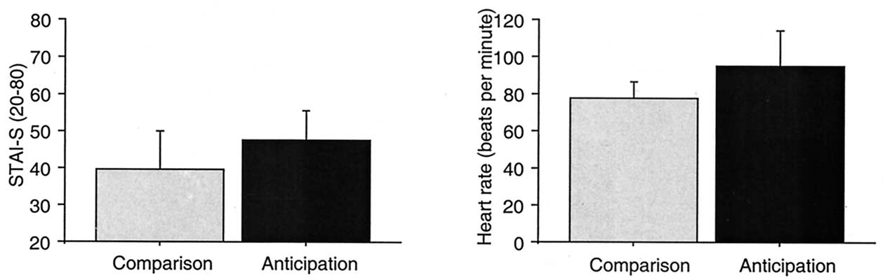 Figure 1. Mean values and standard deviations for subjective measures of state anxiety and heart rate during speaking alone before (i.e.,the anticipation group) or after (i.e., the comparison group) speaking in public for 18 individuals with social phobia.
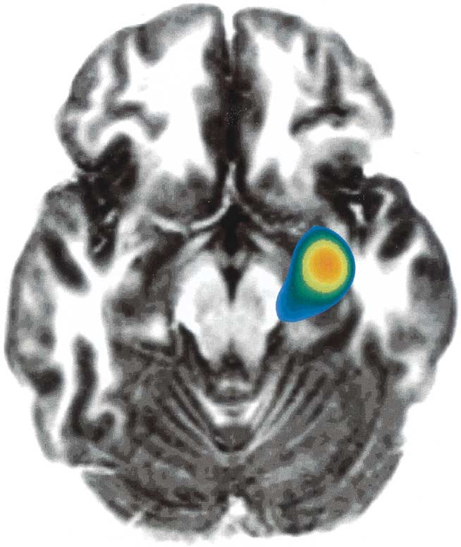 43, z ϭ 32). Thus, task repetition did not influence rCBFin structures associated with anticipatory anxiety except inthe prefrontal cortex where rCBF was attenuated by taskrepetition, but elevated as a function of anticipatoryanxiety. Anticipatory related rCBF-increases were ob-served in the dorsolateral prefrontal cortex while repetitionrelated decreases were located in the prefrontal cortexsuperior to the anticipatory cluster. Because the oppositedirection of change and since the centers of gravity ofthese clusters were separated by 28 mm, it is most likelythat anticipatory anxiety rather than task repetition ac-counts for the observed rCBF alterations in the rightdorsolateral prefrontal cortex.