Scand. J. Lab. Anim. Sci. No. 4. 2004. Vol. 31
Case Report and Short Communication: Rectal prolapse associated with an unusual combination of pin- worms and citrobacter species infection in FVB mice colony
by MJ Mahesh Kumar, P Nagarajan, R Venkatesan & Ramesh C Juyal
Small Animal facility, National Institute of Immunology, New Delhi – 110067, India
Summary Spontaneous cases of rectal prolapse in a breeding colony of FVB mice were found to be due to infection with Syphacia obvelata and Citrobacter freundii. Microbiology, biochemical and parasitological examina- tion revealed Citrobacter freundii and eggs of Syphacia obvelata. After treatment with antibiotics, anti- helminthic drugs and manual reduction prevented further occurrence. Introduction Colitis in mice is associated with a high incidence of
guideline for care and use of animals in scientific
rectal prolapse due to heavy infestation by
research (Indian National Science Academy, New
Citrobacter freundii (Ediger et al., 1974). Rectal
Delhi, India) in a CPCSEA (Committee for the
prolapse due to a natural outbreak of Syphacia
Purpose of Control and Supervision of Experimental
infection (Hoag, 1961; Harwell & Boyd, 1968) and
Animals) registered animal facility. The animals were
due to Citrobacter freundii were reported by Barhold
maintained under the standard environmental condi-
(1980). Other causes for rectal prolapse involved
tion (Temp. 22 C) relative humidity 45-55%) with
pelvic floor disorder in male transgenic mouse
12: 12 hrs dark / light photopheriod and fed commer-
strain deficient in urokinase – plasminogen activator
cial autoclaved pellet feed and water ad libitum. For
(UPA -/-) (Yiou et al., 2001) and in large bowel parasitological examination we used the cellophanedisease caused by Helicobacter hepaticus in
tape method in which cellophane adhesive impres-
immunodeficient mice (Ncr-nu/nu, Balb/ c an Ncr
sions of the anus were applied over the glass slides
nu/ nu, C57 BL / 6 Ncr – nu / nu) (Ward et al, 1996).
and examined under a microscope. In addition, a
Rectal prolapse with colonic mucosal hyperplasia
swab from a rectal prolapse was taken, dissolved in a
was recorded due to citrobacter infection by Barhold
0.1% NaCl solution, centrifuged at 2500 rpm for five
et al. (1977). In this paper we present a case report
minutes, the supernatant solution discarded, and a
describing the rectal prolapse associated with both
drop taken for light microscopic examination.
pinworm and Citrobacter freundii infection in a
Aseptically collected faecal swabs from the pro-
FVB mice colony and its curative measures.
lapsed animals were subjected to culture in blood
agar plates at 37 C for 48 hours under anaerobic
conditions as described (Ediger et al., 1974; Hansen,
In the breeding colony of our small-animal facility,
2000). The pure colonies were subjected to colony
spontaneous rectal prolapses were observed in a
morphology; gram staining reaction (Gram staining
group of the FVB strain. The animals remained kit, Hi Media, India) and biochemical tests were car-housed in individual cages in accordance with the
ried out by a commercial kit for Enterobacteriaceae(HIMVIC Biochemical test Kit KB 003, Hi Media,India). After confirmation the animals were treated
* Correspondance: Small Animal facility, National
with Tetracycline VET water soluble (Intervet, India)
Institute of Immunology, New Delhi – 110067, India.
at a dose rate of 1g / liter of water for three days and
Phone # 91-11-26717128. Fax 91-11-26162125
four consecutive doses of Ivermectin and the pro-
Scand. J. Lab. Anim. Sci. No. 4. 2004. Vol. 31
Results Discussion
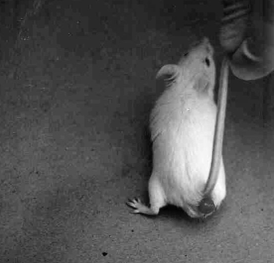
On physical examination, the animals had ruffled
The case describes rectal prolapse associated with
fur and pasty diarrheic faeces around the prolapsed
both pinworm and Citrobacter species infection and
mass, base of the tail and perineum. The mass
their treatment. Heavy infestation of pinworms has
appeared edematous, enlarged with hemorrhagic
previously been reported to cause rectal prolapse,
constipation, intersucception or faecal impactionbut usually without diagnostic procedures beingused to exclude other diseases as primary or con-tributing causes (Wescott et al., 1982). Clinicalsigns associated with pinworms are poor condition,rough hair coats, reduced growth rate and rectalprolapse (Hoag 1961; Harwell & Boyd, 1968). Based on the microscopic examination of faecalsamples, we identified Syphacia species but theclinical signs persisted even after treating with anti-helminthic drugs. Cultural examination followed bya biochemical test for the characterization of thebacteria in the fecal sample revealed the typical
Figure 1: FVB Male mice with Rectal prolapse
colony morphology and biochemical character ofCitrobacter species (Hansen et al., 2000). Hence we
Microscopic examination of faecal samples showed
concluded that the colony was infected with
numerous eggs of Syphacia species. The eggs
Citrobacter bacteria. Based on that, we decided to
appeared asymmetrical, thin shelled, transparent,
treat with an antibiotic and continued with anti-
and slightly flattened on one side and resembled a
helminthics. While antihelminthics are effective in
eliminating a high percentage of adult pinworm,
Culture examination revealed typical large round
these are inefficient in clearing immature worms
colonies with a smooth edge surface. Gram staining
(Wescot et al., 1982). The affected animals returned
reaction of pure colonies showed multiple numbers
to normal after five days of treatment.
of gram-negative bacteria that were pale in colorwith pleomorphic rod-shaped organisms. Acknowledgement
Biochemical examination of pure colonies using a
We are thankful to the Director NII for giving nec-
commercial kit showed positive for sucrose, H S
essary support to carry out the study.
and negative for lysine, ornithine decarboxylaseand the VP test. Based on these results we decided
References:
to treat the animal with tetracycline hydrochloride 1
Barthold SW, OL Osbaldistob & AM Jonas: Dietary
gm / liter of water for 3 days and Ivermectin 4 mg/
bacterial and host genetic interaction in the
kg body weight (Klement et al., 1996) for four con-
pathogensis of transmissible murine colonic
secutive days. After treatment, all clinical signs dis-
hyperplasia. Laboratory animal science. 1977,
appeared except the prolapsed mass. The prolapsed
mass was cleaned with normal saline and reduced
Barthold SW: The microbiology of transmissible
manually; following reduction there was no recur-
murine colonic hyperplasia. Laboratory animal
Scand. J. Lab. Anim. Sci. No. 4. 2004. Vol. 31
Ediger RD, RM Kovatch & MM Rabstain: Colitis in
tory rats and mice. Laboratory Animal Science.
mice with a high incidence of rectal prolapse.
Laboratory animal science. 1974, 24, 488- 494. Ward JM, MR Anver, DC Haines, JM Melhorn, PHansen AK.: Handbook of laboratory animal bacte-
Gorelick, P & JG Yanl: Inflammatory large
riology. CRC Press, Washington, D.C. (2000),
bowel disease in immuno deficient mice natu-
rally infected with helicobacter hepaticus. Harwell JF & DD Boyd: Naturally occurring
Laboratory Animal Science. 1996, 46 (1) 15-20.
oxyuris in mice. Journal of the American veteri-
Wescott RB: Helminths. In: The mouse in biomed-
nary medical association. 1968, 153, 950 – 953.
ical research, New York: Academic Press, 1982,
Hoag WG: Oxyuriasis in laboratory mouse
Yiou R, U Delmas & P Carmeliet: The pathophysi-
Research. 1961, 22, 150 –153.
ology of pelvic floor disorders. Evidence from
Klement P, JM Augustine, KH Delaney, G Klement
histomorphologic study of the perineum and a
& JL Weitz: An oral ivermectin regimen that
mouse model of rectal prolapse. Journal of
eradicates pinworms (Syphacia spp.) in labora-
Anatomy. 2001, 199, 599 – 607.
GEGEN JEDES ÜBEL IST EIN KRAUT GEWACHSEN Krankheitsbilder und eine Auswahl natürlicher Heilmittel Wählen sie intuitiv, auf welches Heilkraut sie ansprechen! Alant, Apfel, Faulbaum, Hafer, Knoblauch, Lein, Meerrettich, PaprikaApfel, Baldrian, Hafer, Holunder, Johanniskraut, Kamille, MelisseBrennessel, Hagebutten, Sanddorn, Schafgarbe, Tausendgulden-kraut, Wacholder, Weinrebe, Weizen,
Was Sie selbst tun sollten Was Partner tun können Gehen Sie zunächst zum Hausarzt – er wird die grundlegenden Seien Sie verständnisvoll und geben Sie Mut. Faktoren abklären und Sie bei Bedarf zum Urologen überweisen. Üben Sie keinen Druck aus, haben Sie Scheuen Sie nicht den Weg zum Urologen; nehmen Sie dort Partner, einen Arzt ins Vertrauen zu ziehen. Zi
 Scand. J. Lab. Anim. Sci. No. 4. 2004. Vol. 31
Results
Scand. J. Lab. Anim. Sci. No. 4. 2004. Vol. 31
Results